
ICD-9-CM 362.50 MedlinePlus 001000 | ICD-10 H35.3 DiseasesDB 11948 eMedicine article/1223154 | |
 | ||
Macular degeneration, also known as age-related macular degeneration (AMD or ARMD), is a medical condition which may result in blurred or no vision in the center of the visual field. Early on there are often no symptoms. Over time, however, some people experience a gradual worsening of vision that may affect one or both eyes. While it does not result in complete blindness, loss of central vision can make it hard to recognize faces, drive, read, or perform other activities of daily life. Visual hallucinations may also occur but these do not represent a mental illness.
Contents
- Signs and symptoms
- Risk factors
- Environment and lifestyle
- Genetics
- Specific genes
- Mitochondrial related gene polymorphisms
- Pathophysiology
- Stages
- Early AMD
- Intermediate AMD
- Late AMD
- Geographic Atrophy
- Oxidative stress
- Diagnosis
- Histology
- Prevention
- Management
- Dry AMD
- Wet AMD
- Adaptive devices
- Epidemiology
- Association with other age related diseases
- Genetic testing
- Stem cell transplant
- Other types
- Notable cases
- References
Macular degeneration typically occurs in older people. Genetic factors and smoking also play a role. It is due to damage to the macula of the retina. Diagnosis is by a complete eye exam. The severity is divided into early, intermediate, and late types. The late type is additionally divided into "dry" and "wet" forms with the dry form making up 90% of cases.
Prevention includes exercising, eating well, and not smoking. Antioxidant vitamins and minerals do not appear to be useful for prevention. There is no cure or treatment that returns vision already lost. In the wet form, anti-VEGF medication injected into the eye or less commonly laser coagulation or photodynamic therapy may slow worsening. Supplements in those who already have the disease may slow progression.
In 2010 it affected 23.5 million people globally. In 2013 moderate to severe disease affected 13.4 million and it is the fourth most common cause of blindness after cataracts, preterm birth, and glaucoma. It most commonly occurs in people over the age of fifty and in the United States is the most common cause of vision loss in this age group. About 0.4% of people between 50 and 60 have the disease, while it occurs in 0.7% of people 60 to 70, 2.3% of those 70 to 80, and nearly 12% of people over 80 years old.
Signs and symptoms
Signs and symptoms of macular degeneration include:
Macular degeneration by itself will not lead to total blindness. For that matter, only a very small number of people with visual impairment are totally blind. In almost all cases, some vision remains, mainly peripheral. Other complicating conditions may possibly lead to such an acute condition (severe stroke or trauma, untreated glaucoma, etc.), but few macular degeneration patients experience total visual loss.
The area of the macula comprises only about 2.1% of the retina, and the remaining 97.9% (the peripheral field) remains unaffected by the disease. Even though the macula provides such a small fraction of the visual field, almost half of the visual cortex is devoted to processing macular information.
The loss of central vision profoundly affects visual functioning. It is quite difficult, for example, to read without central vision. Pictures that attempt to depict the central visual loss of macular degeneration with a black spot do not really do justice to the devastating nature of the visual loss. This can be demonstrated by printing letters six inches high on a piece of paper and attempting to identify them while looking straight ahead and holding the paper slightly to the side. Most people find this difficult to do.
Risk factors
Environment and lifestyle
Genetics
Recurrence ratios for siblings of an affected individual are three- to sixfold higher than in the general population. Genetic linkage analysis has identified 5 sets of gene variants at three locations on different chromosomes (1, 6 and 10) as explaining at least 50% of the risk. These genes have roles regulating immune response, inflammatory processes and homeostasis of the retina. Variants of these genes give rise to different kinds of dysfunction in these processes. Over time, this results in accumulation of intracellular and extracellular metabolic debris. This can cause scarring of the retina or breakdown of its vascularization.
Genetic tests are available for some of these gene variations. However, pathogenesis of macular degeneration is a complex interaction between genetics, environment and lifestyle, and presence of unfavorable genetic factors doesn't necessarily predict progression to disease. The three loci where identified gene variants are found are designated:
Specific genes
Mitochondrial related gene polymorphisms
such as that in the MT-ND2 molecule, predicts wet AMD.
Pathophysiology
The pathogenesis of age-related macular degeneration is not well known, although a number of theories have been put forward, including oxidative stress, mitochondrial dysfunction, and inflammatory processes.
The imbalance between production of damaged cellular components and degradation leads to the accumulation of detrimental products, for example, intracellular lipofuscin and extracellular drusen. Incipient atrophy is demarcated by areas of retinal pigment epithelium (RPE) thinning or depigmentation that precede geographic atrophy in the early stages of AMD. In advanced stages of AMD, atrophy of the RPE (geographic atrophy) and/or development of new blood vessels (neovascularization) result in death of photoreceptors and central vision loss.
In the dry (nonexudative) form, cellular debris called drusen accumulates between the retina and the choroid, causing atrophy and scarring to the retina. In the wet (exudative) form, which is more severe, blood vessels grow up from the choroid (neovascularization) behind the retina which can leak exudate and fluid and also cause hemorrhaging.
Early work demonstrated a family of immune mediators was plentiful in drusen. Complement factor H (CFH) is an important inhibitor of this inflammatory cascade, and a disease-associated polymorphism in the CFH gene strongly associates with AMD. Thus an AMD pathophysiological model of chronic low grade complement activation and inflammation in the macula has been advanced. Lending credibility to this has been the discovery of disease-associated genetic polymorphisms in other elements of the complement cascade including complement component 3 (C3).
A powerful predictor of AMD is found on chromosome 10q26 at LOC 387715. An insertion/deletion polymorphism at this site reduces expression of the ARMS2 gene though destabilization of its mRNA through deletion of the polyadenylation signal. ARMS2 protein may localize to the mitochondria and participate in energy metabolism, though much remains to be discovered about its function.
Other gene markers of progression risk includes tissue inhibitor of metalloproteinase 3 (TIMP3), suggesting a role for intracellular matrix metabolism in AMD progression. Variations in cholesterol metabolising genes such as the hepatic lipase, cholesterol ester transferase, lipoprotein lipase and the ABC-binding cassette A1 correlate with disease progression. The early stigmata of disease, drusen, are rich in cholesterol, offering face validity to the results of genome-wide association studies.
Stages
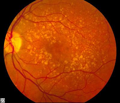
In AMD there is a progressive accumulation of characteristic yellow deposits, called drusen (buildup of extracellular proteins and lipids), in the macula (a part of the retina), between the retinal pigment epithelium and the underlying choroid which is believed to damage the retina over time. Amyloid beta, which builds up in Alzheimer's disease brains, is one the proteins accumulating in AMD, which is one of the reasons AMD is sometimes called "Alzheimer's of the eye" or "Alzheimer's of the retina". AMD can be divided into 3 stages: early, intermediate, and late, based partially on the extent (size and number) of drusen.
AMD-like pathology begins with small yellow deposits (drusen) in the macula, between the retinal pigment epithelium and the underlying choroid. Most people with these early changes (referred to as age-related maculopathy) still have good vision. People with drusen may or may not develop AMD, in fact the majority of people over age 60 have drusen with no negative effects. The risk of developing symptoms is higher when the drusen are large and numerous and associated with disturbance in the pigmented cell layer under the macula. Large and soft drusen are thought to be related to elevated cholesterol deposits.
Early AMD
Early AMD is diagnosed based on the presence of medium-sized drusen, about the width of an average human hair. Early AMD is usually asymptomatic.
Intermediate AMD
Intermediate AMD is diagnosed by large drusen and/or any retinal pigment abnormalities. Intermediate AMD may cause some vision loss, however, like Early AMD, it is usually asymptomatic.
Late AMD
In late AMD, enough retinal damage occurs that people have symptomatic central vision loss in addition to drusen. The damage can either be development of atrophy or the onset of neovascular disease. Late AMD is further divided into two subtypes based on the types of damage: Geographic atrophy and Wet AMD (also called Neovascular AMD).
Dry AMD
Dry AMD (also called nonexudative AMD) is a broad designation, encompassing all forms of AMD that are not neovascular (wet AMD). This includes early and intermediate forms of AMD, as well as the advanced form of dry AMD known as geographic atrophy. Dry AMD patients tend to have minimal symptoms in the earlier stages; visual function loss occurs more often if the condition advances to geographic atrophy. Dry AMD accounts for 80–90% of cases and tend to progress slowly. In 10–20% of people, dry AMD progresses to the wet type.
Geographic Atrophy
Geographic atrophy (also called atrophic AMD) is an advanced form of AMD in which progressive and irreversible loss of retinal cells leads to a loss of visual function.
Wet AMD
Neovascular or exudative AMD, the "wet" form of advanced AMD, causes vision loss due to abnormal blood vessel growth (choroidal neovascularization) in the choriocapillaris, through Bruch's membrane. It is usually, but not always, preceded by the dry form of AMD. The proliferation of abnormal blood vessels in the retina is stimulated by vascular endothelial growth factor (VEGF). Unfortunately, because these blood vessels are abnormal, these new vessels are fragile, ultimately leading to blood and protein leakage below the macula. Bleeding, leaking, and scarring from these blood vessels eventually cause irreversible damage to the photoreceptors and rapid vision loss if left untreated.
Oxidative stress
Age-related accumulation of low-molecular-weight, phototoxic, pro-oxidant melanin oligomers within lysosomes in the retinal pigment epithelium (RPE) may be partly responsible for decreasing the digestive rate of photoreceptor outer rod segments (POS) by the RPE – autophagy. A decrease in the digestive rate of POS has been shown to be associated with lipofuscin formation – a classic sign associated with AMD.
The role of retinal oxidative stress in the etiology of AMD by causing further inflammation of the macula is suggested by the enhanced rate of disease in smokers and those exposed to UV irradiation.
Mitochondrial dysfunction may play a role.
Diagnosis
Diagnosis of age-related macular degeneration rests on signs in the macula, irrespective of visual acuity. Diagnosis of AMD may include the following procedures and tests:
Histology
Prevention
A 2012 Cochrane review found the use of vitamin and mineral supplements, alone or in combination, by the general population had no effect on whether or not AMD started.
Management
Supplements that include lutein and zeaxanthin may slow down the worsening of AMD. They have; however, not been shown to prevent the disease. There is not enough evidence to determine if statins have a role in preventing or slowing the progression of AMD. Antiangiogenic steroids such as anecortave acetate and triamcinolone acetonide have shown no evidence in preventing visual loss in people with neovascular AMD.
Dry AMD
No medical or surgical treatment is available for this condition.
Wet AMD
It can be treated with laser coagulation, and more commonly with medication that stops and sometimes reverses the growth of blood vessels.
A randomized control trial found that bevacizumab and ranibizumab had similar efficacy, and reported no significant increase in adverse events with bevacizumab. A 2014 Cochrane review found that the systemic safety of bevacizumab and ranibizumab are similar when used to treat neovascular AMD, except for gastrointestinal disorders. Bevacizumab however is not FDA approved for treatment of macular degeneration. A controversy in the UK involved the off-label use of cheaper bevacizumab over the approved, but expensive, ranibizumab. Ranibizumab is a smaller fragment, Fab fragment, of the parent bevacizumab molecule specifically designed for eye injections. Other approved antiangiogenic drugs for the treatment of neo-vascular AMD include pegaptanib and aflibercept.
The American Academy of Ophthalmology practice guidelines do not recommend laser coagulation therapy for macular degeneration, but state that it may be useful in people with new blood vessels in the choroid outside of the fovea who don't respond to drug treatment. There is strong evidence that laser coagulation will result in the disappearance of drusen but does not affect choroidal neovascularisation. A 2007 Cochrane review on found that laser photocoagulation of new blood vessels in the choroid outside of the fovea is effective and economical method, but that the benefits are limited for vessels next to or below the fovea.
Photodynamic therapy has also been used to treat wet AMD. The drug verteporfin is administered intravenously; light of a certain wavelength is then applied to the abnormal blood vessels. This activates the verteporfin destroying the vessels.
Cataract surgery could possibly improve visual outcomes for people with AMD, though there have been concerns of surgery increasing the progression of AMD. A randomized controlled trial found that people who underwent immediate cataract surgery (within 2 weeks) had improved visual acuity and better quality of life outcomes than those who underwent delayed cataract surgery (6 months).
Adaptive devices
Because peripheral vision is not affected, people with macular degeneration can learn to use their remaining vision to partially compensate. Assistance and resources are available in many countries and every state in the U.S. Classes for "independent living" are given and some technology can be obtained from a state department of rehabilitation.
Adaptive devices can help people read. These include magnifying glasses, special eyeglass lenses, computer screen readers, and TV systems that enlarge reading material.
Computer screen readers such as JAWS or Thunder work with standard Windows computers. Also Apple devices provide wide range of features (voice over,screen readers, Braille etc.,
Video cameras can be fed into standard or special-purpose computer monitors, and the image can be zoomed in and magnified. These systems often include a movable table to move the written material.
Accessible publishing provides larger fonts for printed books, patterns to make tracking easier, audiobooks and DAISY books with both text and audio.
Epidemiology
Age-related macular degeneration accounts for more than 54% of all vision loss in the white population in the USA. An estimated 8 million Americans are affected with early age-related macular degeneration, of whom over 1 million will develop advanced age-related macular degeneration within the next 5 years. In the UK, age-related macular degeneration is the cause of blindness in almost 42% of those who go blind aged 65–74 years, almost two-thirds of those aged 75–84 years, and almost three-quarters of those aged 85 years or older.
Macular degeneration is more likely to be found in Caucasians than in people of African descent.
Association with other age-related diseases
Studies indicate drusen associated with AMD are similar in molecular composition to Beta-Amyloid (βA) plaques and deposits in other age-related diseases such as Alzheimer's disease and atherosclerosis. This suggests that similar pathways may be involved in the etiologies of AMD and other age-related diseases.
Genetic testing
A practical application of AMD-associated genetic markers is in the prediction of progression of AMD from early stages of the disease to neovascularization.
Stem cell transplant
Cell based therapies using bone marrow stem cells as well as Retinal pigment epithelial transplantation are being studied. Recent advancements within the field of stem cell research in the United States have led to the first human embryonic stem cell trial for dry AMD, which reports positive results.
Other types
There are a few other (rare) kinds of macular degeneration with similar symptoms but unrelated in etiology to Wet or Dry age-related macular degeneration. They are all genetic disorders that may occur in childhood or middle age.
Similar symptoms with a very different etiology and different treatment can be caused by epiretinal membrane or macular pucker or any other condition affecting the macula, such as central serous retinopathy.