
ICD-9-CM 95.12 | MeSH D005451 | |
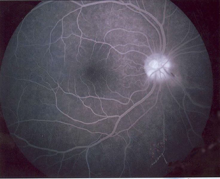 | ||
Fluorescein angiography (FA), fluorescent angiography (FAG), or fundus fluorescein angiography (FFA) is a technique for examining the circulation of the retina and choroid (parts of the fundus) using a fluorescent dye and a specialized angiographic camera. It involves administration of sodium fluorescein into the systemic circulation, and then an angiogram is obtained by photographing the fluorescence emitted after illumination of the retina with blue light at a wavelength of 490 nanometers. The test uses the dye tracing method. The fluorescein is administered intravenously in intravenous fluorescein angiography (IVFA) and orally in oral fluorescein angiography (OFA).
Contents
- Equipment
- Technique
- Normal circulatory filling
- Pathologic findings
- Other types of fluorescent angiography
- References
The fluorescein dye also reappears in the patient urine, causing the urine to appear darker, and sometimes orange. It can also cause discolouration of the saliva.
Fluorescein angiography is one of several health care applications of this dye, all of which have a risk of severe adverse effects. See fluorescein safety in health care applications. Fluorescein angiography does not involve the use of ionizing radiation.
Equipment
Technique
Normal circulatory filling
times are approximate
Fluorescein enters the ocular circulation from the internal carotid artery via the ophthalmic artery. The ophthalmic artery supplies the choroid via the short posterior ciliary arteries and the retina via the central retinal artery, however, the route to the choroid is typically less circuitous than the route to the retina. This accounts for the short delay between the "choroidal flush" and retinal filling.
Pathologic findings
Pathologic changes are recognized by the detection of either hyperfluorescence or hypofluroescence.
Causes of hyperfluorescence:
window/transmission (filling) defectsleaking defects (i.e. capillary leakage, aneurysm, neovascularization)pooling defectsstainingabnormal vasculatureCauses of hypofluorescence:
blocking defect (i.e. blood)filling defect (capillary nonperfusion/blockage)Fluorescein angiography is used by physicians specializing in the treatment of eye diseases (ophthalmologists) to evaluate the vasculature of the retina, choroid, optic disc, and iris. Among the common groups of ophthalmologic disease, fluorescein angiography can detect diabetic retinopathy (neovascularization), vein occlusions, retinal artery occlusions, edema of the optic disc, and tumors. Additionally, the transit time (the period between injection of the dye and when it appears in the examined blood vessels) can provide an objective measurement of the rate of blood flow through the imaged blood vessels.