The infratemporal fossa is an irregularly shaped cavity, situated below and medial to the zygomatic arch. It is not fully enclosed by bone in all directions, and it contains superficial muscles that are visible during dissection after removing skin and fascia: namely, the lower part of the temporalis muscle, the lateral pterygoid, and the medial pterygoid.
Its boundaries may be defined by:
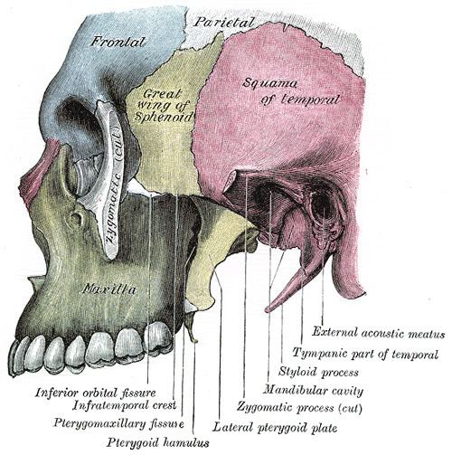
anteriorly, by the infratemporal surface of the maxilla and the ridge which descends from its zygomatic processposteriorly, by the articular tubercle of the temporal and the spina angularis of the sphenoidsuperiorly, by the greater wing of the sphenoid below the infratemporal crest, and by the under surface of the temporal squama, containing the foramen ovale, which transmits the mandibular branch of the trigeminal nerve, and the foramen spinosum, which transmits the middle meningeal arteryinferiorly, by the medial pterygoid muscle attaching to the mandiblemedially, by the lateral pterygoid platelaterally, by the ramus of mandible, which contains the mandibular foramen, leading to the mandibular canal through which the inferior alveolar nerve passes. This also contains the lingula, a triangular piece of bone that overlies the mandibular foramen antero-medially. Finally, the mylohyoid groove descends obliquely transmitting the mylohyoid nerve the only motor branch of the posterior division of the trigeminal nerve.Lower part of the Temporalis and masseter muscles (origin of masseter muscle:lower margin of the inner surface of zygomatic bone insertion : outer surface of the ramus of the mandible )Lateral and medial pterygoid musclesThe internal maxillary vessels, consisting of the maxillary artery originating from the external carotid artery and its branches.
Internal maxillary branches found within the infratemporal fossa including the
middle meningeal arteryinferior alveolar arterydeep temporal arterybuccal arterypterygoid venous plexusretromandibular veinMandibular nerve, inferior alveolar nerve, lingual nerve, buccal nerve, chorda tympani nerve, and otic ganglion.
Mandibular nerve
Mandibular nerve which is the third branch of the trigeminal nerve (CN V3), also known as the "inferior maxillary nerve" or nervus mandibularis, enters infratemporal fossa from middle cranial fossa through foramen ovale.Motor branches:
masseteric nervedeep temporal nervelateral pterygoid nerve and medial pterygoid nerveIts motor fibers innervate all the muscles of mastication plus the mylohyoid, anterior belly of the digastric, and the tensores veli palati and tympani
Sensory innervation:
meningeal nervebuccal nerveauriculotemporal nervelingual nerveinferior alveolar nerveauricleexternal acoustic meatustympanic membranetemporal regioncheekskin overlying the mandible (except at the angle of the mandible)floor of mouthlower teethgingivaCranial cavity (through foramen ovale and spinosum).Temporal fossa (deep to zygomatic arch).pterygopalatine fossa (through pterygomaxillary fissure).Orbit (through inferior orbital fissure).parapharyngeal space.The foramen ovale and foramen spinosum open on its roof, and the alveolar canals on its anterior wall.
At its upper and medial part are two fissures, which together form a T-shaped fissure, the horizontal limb being named the inferior orbital, and the vertical one the pterygomaxillary.