
Artery masseteric artery Latin musculas masseter | Nerve mandibular nerve (V3) | |
 | ||
Actions elevation (as in closing of the mouth) and protraction of mandible | ||
In human anatomy, the masseter is one of the muscles of mastication. In the animal kingdom, it is particularly powerful in herbivores to facilitate chewing of plant matter. The most obvious muscle of mastication is the masseter muscle, since it is the most superficial and one of the strongest.
Contents
Structure
The masseter is a thick, somewhat quadrilateral muscle, consisting of two heads, superficial and deep. The fibers of the two heads are continuous at their insertion.
Superficial head
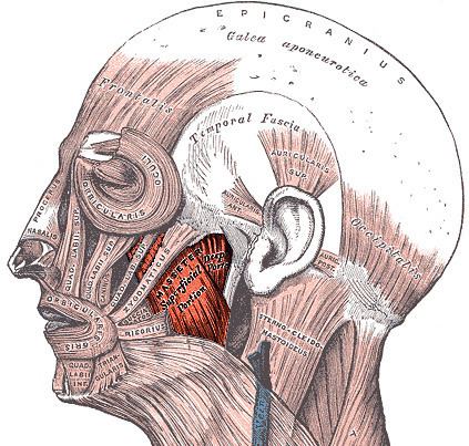
The superficial head, the larger, arises by a thick, tendinous aponeurosis from the maxillary process of the zygomatic bone, and from the anterior two-thirds of the inferior border of the zygomatic arch. Its fibers pass inferior and posterior, to be inserted into the angle of the mandible and inferior half of the lateral surface of the ramus of the mandible.
Deep head
The deep head is much smaller,and more muscular in texture. It arises from the posterior third of the lower border and from the whole of the medial surface of the zygomatic arch. Its fibers pass downward and forward, to be inserted into the upper half of the ramus as high as the coronoid process of the mandible. The deep head of the muscle is partly concealed, anteriorly, by the superficial portion. Posteriorly, it is covered by the parotid gland.
Innervation
Along with the other three muscles of mastication (temporalis, medial pterygoid, and lateral pterygoid), the masseter is innervated by the anterior division of the mandibular division (V3) of the trigeminal nerve. The innervation pathway is: gyrus precentralis > genu capsula interna > nucleus motorius nervi trigemini > nervus trigeminus > nervus mandibularis > musculus masseter.
Function
The action of the muscle during bilateral contraction of the entire muscle is to elevate the mandible, raising the lower jaw. Elevation of the mandible occurs during the closing of the jaws. The masseter parallels the medial pterygoid muscle, but it is stronger.
Examination
During an extraoral examination, stand near the patient and visually inspect and bilaterally palpate the muscle. Place the fingers of each hand over the muscle and ask the patient to clench his or her teeth several times.
Pathology
The masseter muscle can become enlarged in patients who habitually clench or grind (with bruxism) their teeth and even in those who constantly chew gum. This masseteric hypertrophy is asymptomatic and soft; it is usually bilateral but can be unilateral. Even if the hypertrophy is bilateral, asymmetry of the face may still occur due to unequal enlargement of the muscles. This extraoral enlargement may be confused with parotid salivary gland disease, dental infections, and maxillofacial neoplasms. However, no other signs are present except those involved in changes in occlusion intraorally such as pain, and the enlargement corresponds with the outline of the muscle. Most patients seek medical attention because of comments about facial appearance, and this situation may be associated with further pathology of the temporomandibular joint.
Finally, the muscle undergoes spasm with malignant hyperthermia as do other skeletal muscles, but this one is easily noted, since it is on the face.