Suvarna Garge (Editor)

Herpes zoster ophthalmicus

Updated on
Edit
Like
Comment
Share on FacebookTweet on TwitterShare on LinkedInShare on Reddit
Specialty
  
infectious disease

ICD-9-CM
  
053.20

MedlinePlus
  
000858

ICD-10
  
B02

DiseasesDB
  
29119

Herpes zoster ophthalmicus

eMedicine
  
med/1007 derm/180 emerg/823 oph/257 ped/996

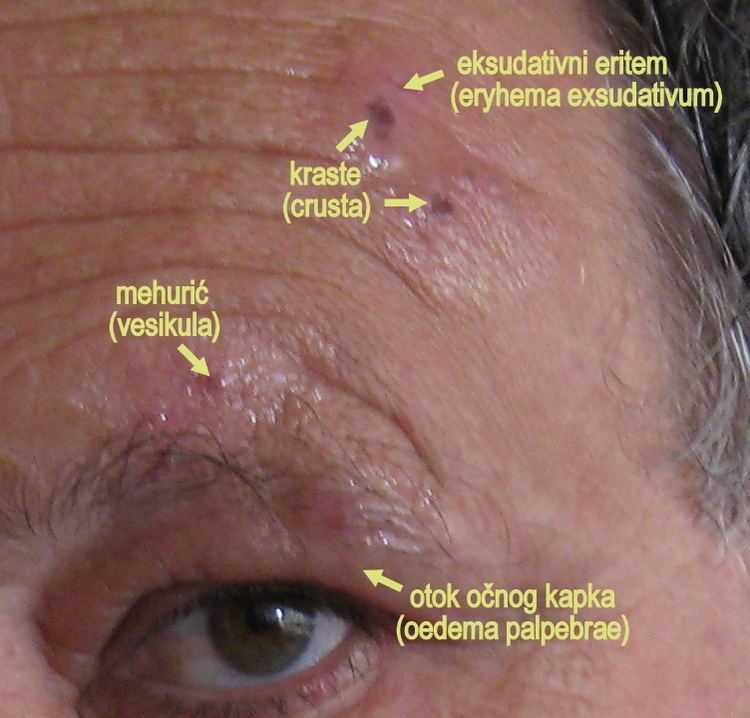
Herpes zoster ophthalmicus (HZO) and also known as ophthalmic zoster is a disease characterised by reactivation of dormant varicella zoster virus residing within the ophthalmic nerve (the first division of the trigeminal nerve). This condition is an important subtype of shingles, representing 15% of all cases. Herpes zoster ophthalmicus is transmitted via direct contact or droplets. Varicella zoster virus is a DNA virus which produces acidophilic intranuclear inclusion bodies. The virus is neurotrophic in nature.

Contents

The frontal nerve is more commonly affected than the nasociliary nerve or lacrimal nerve.

Signs and symptoms

Any potential ocular involvement should be assessed by an ophthalmologist as complications such as episcleritis and uveitis may occur.

Skin

  • Viral prodrome
  • Preherpetic neuralgia
  • Rash, transitioning from papules to vesicles to pustules to scabs.
  • Hutchinson's sign: cutaneous involvement of the tip of the nose, indicating nasociliary nerve involvement. A positive Hutchinson's sign increases the likelihood of ocular complications associated with HZO.
  • Disseminated distribution in individuals with immunodeficiency.
  • Cornea

  • Epithelial: punctate epithelial erosions and pseudodendrites: often have anterior stromal infiltrates. Onset 2 to 3 days after the onset of the rash, resolving within 2–3 weeks. Common.
  • Stromal:
  • Nummular keratitis: have anterior stromal granular deposits. Occurs within 10 days of onset of rash. Uncommon
  • Necrotising interstitial keratitis: Characterised by stromal infiltrates, corneal thinning and possibly perforation. Occurs between 3 months and several years after onset of rash. Rare.
  • Disciform Keratitis(Disciform Endotheliitis): a disc of corneal oedema, folds in Descemet's membrane, mild inflammation evident within the anterior chamber and fine keratic precipitates. Chronic. Occurs between 3 months and several years after the onset of the rash. Uncommon.
  • Neurotrophic: corneal nerve damage causes persistent epithelial defect, thinning and even perforation. Cornea becomes susceptible to bacterial and fungal keratitis. Chronic. Late onset. Uncommon.
  • Mucous plaques: linear grey elevations loosely adherent to the underlying diseased epithelium/stroma. Chronic. Onset between 3 months and several years after the onset of the rash.
  • Uveal

    Anterior uveitis develops in 40–50% of cases with HZO within 2 weeks of onset of the skin rashes. Typical HZO keratitis at least mild iritis, especially if Hutchinson's sign is positive for the presence of vescicles upon the tip of the nose.

    Features:

    This non-granulomatous iridocyclitis is associated with:

  • Small keratic precipitates
  • Mild aqueous flare
  • Occasionally haemorrhagic hypopion.
  • HZO uveitis is associated with complications such as iris atrophy and secondary glaucoma are not uncommon. Complicated cataract may develop in the late stages of the disease.

    Treatment

    Treatment is usually with antivirals such as aciclovir orally.

    Uveal

  • Topical steroids and cycloplegics for several months.
  • Systemic Aciclovir facilitates the early control of HZO lesions.
  • References

    Herpes zoster ophthalmicus Wikipedia