
Dorlands/Elsevier n_05/12566272 FMA 52668 | Latin nervus nasociliaris TA A14.2.01.025 | |
 | ||
To long root of the ciliary ganglion, the long ciliary nerves, the infratrochlear nerve, and the ethmoidal nerves | ||
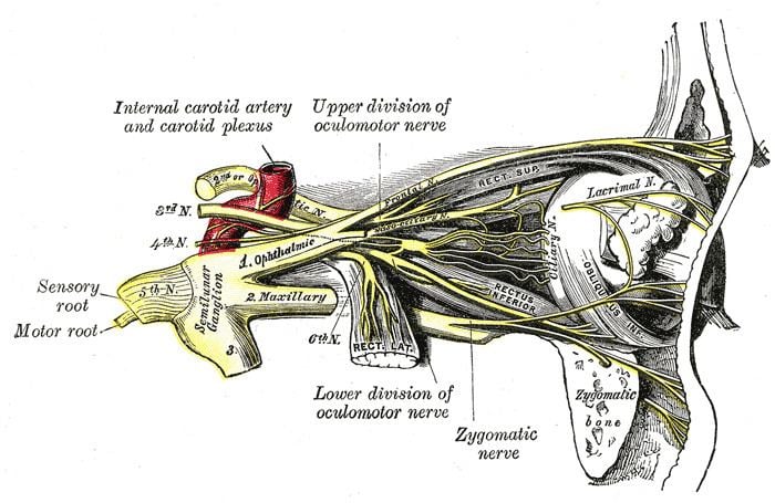
The nasociliary nerve is a branch of the ophthalmic nerve (CN V1; one of three branches of the trigeminal nerve a.k.a. CN V). It is intermediate in size between the two other main branches of the ophthalmic nerve, the frontal nerve and the lacrimal nerve, and is more deeply placed.
Contents
Structure
The nasociliary nerve enters the orbit between the two heads of the lateral rectus muscles and between the superior and inferior rami of the oculomotor nerve (CN III). It passes across the optic nerve (CN II) and runs obliquely beneath the superior rectus muscle and superior oblique muscle to the medial wall of the orbital cavity. It passes through the anterior ethmoidal opening as the anterior ethmoidal nerve and enters the cranial cavity just above the cribriform plate of the ethmoid bone. It supplies branches to the mucous membrane of the nasal cavity and finally emerges between the inferior border of the nasal bone and the side nasal cartilages as the external nasal branch.
Function
A branch of the ophthalmic nerve [CN V1] in the superior orbital fissure, passing through the orbit, giving rise to the communicating branch to the ciliary ganglion, the long ciliary nerves, the posterior and anterior ethmoidal nerves, and terminating as the infratrochlear and nasal branches, which supply the mucous membrane of the nose, the skin of the tip of the nose, and the conjunctiva.
Branches
The nasociliary nerve gives off the following branches:
PLICA is a mnemonic often used to remember these branches.
Testing
Since both the short and long ciliary nerves carry the afferent limb of the corneal blink reflex, one can test the integrity of the nasociliary nerve (and, ultimately, the trigeminal nerve) by examining this reflex in the patient. Normally both eyes should blink when either cornea (not the conjunctiva, which is supplied by the adjacent cutaneous nerves) is irritated. If neither eye blinks, then either the ipsilateral nasociliary nerve is damaged, or the facial nerve (CN VII, which carries the efferent limb of this reflex) is bilaterally damaged. If only the contralateral eye blinks, then the ipsilateral facial nerve is damaged. If only the ipsilateral eye blinks, then the contralateral facial nerve is damaged.