
ICD-9-CM 288.4 eMedicine ped/745 | ICD-10 D76.1 DiseasesDB 31418 | |
 | ||
OMIM 267700 603553 608898 603552 | ||
Hemophagocytic lymphohistiocytosis (HLH), also known as haemophagocytic lymphohistiocytosis (British spelling), and hemophagocytic or haemophagocytic syndrome, is an uncommon hematologic disorder. It is a life-threatening disease of severe hyperinflammation caused by uncontrolled proliferation of activated lymphocytes and macrophages, characterised by proliferation of morphologically benign lymphocytes and macrophages that secrete high amounts of inflammatory cytokines. It is classified as one of the cytokine storm syndromes.
Contents
History
The first case report of HLH was published in 1952.
Classification
Primary HLH, also known as familial haemophagocytic lymphohistiocytosis (FHL) or familial erythrophagocytic lymphohistiocytosis, is a heterogeneous autosomal recessive disorder found to be more prevalent with parental consanguinity.
Secondary haemophagocytic lymphohistiocytosis (acquired haemophagocytic lymphohistiocytosis) occurs after strong immunologic activation, such as that which can occur with systemic infection, immunodeficiency, or underlying malignancy.
Both forms are characterized by the overwhelming activation of normal T lymphocytes and macrophages, invariably leading to clinical and haematologic alterations and death in the absence of treatment.
Genetics
Five genetic subtypes (FHL1, FHL2, FHL3, FHL4, and FHL5) are described, with an estimated prevalence of one in 50,000 and equal gender distribution. Molecular genetic testing for four of the causative genes, PRF1 (FHL2), UNC13D (FHL3), STX11 (FHL4), and STXBP2 (FHL5), is available on a clinical basis. Symptoms of FHL are usually evident within the first few months of life and may even develop in utero. However, symptomatic presentation throughout childhood and even into young adulthood has been observed in some cases.
The five subtypes of FHL are each associated with a specific gene:
Nearly half of the cases of type 2 familial hemophagocytic lymphohistiocytosis are due to bi-allelic PRF1 mutations.
Clinical presentation
The onset of HLH occurs under the age of 1 year in ~70% of cases. Familial HLH should be suspected if siblings are diagnosed with HLH or if symptoms recur when therapy has been stopped. Each full sibling of a child with familial HLH has a 25% chance of developing the disease, a 50% chance of carrying the defective gene (which is very rarely associated with any risk of disease) and a 25% chance of not being affected and not carrying the gene defect.
Patients with HLH, especially when untreated, may need intensive therapy. Therefore, HLH should be included in the differential diagnosis of ICU (Intensive Care Unit) patients with cytopenia and hyperferritinemia.
HLH clinically manifests with fever, hepatosplenomegaly, lymphadenopathy, jaundice and a rash.
Investigations
The blood count typically shows pancytopenia—anemia, neutropenia, and thrombocytopenia.
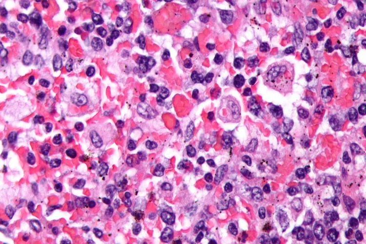
The bone marrow may show hemophagocytosis.
The liver function tests are usually elevated. Hypoalbuminemia is common.
The serum C reactive protein, erythrocyte sedimentation rate, and ferritin level are markedly elevated. In pediatric populations a ferritin above 10000 is very sensitive and specific for the diagnosis of HLH, however, the diagnostic utility for ferritin is less for adult HLH patients.
The serum fibrinogen level is usually low and the D-dimer level is elevated.
The sphingomyelinase is elevated.
Bone marrow biopsy shows histiocytosis.
Diagnostic criteria
The current (2008) diagnostic criteria for HLH are
1. A molecular diagnosis consistent with HLH. These include the identification of pathologic mutations of PRF1, UNC13D, or STX11.
OR
2. Fulfillment of five out of the eight criteria below:
In addition, in the case of familial HLH, no evidence of malignancy should be apparent.
It should be noted that not all five out of eight criteria are required for diagnosis of HLH in adults, and a high index of suspicion is required for diagnosis as delays results in increased mortality. The diagnostic criteria were developed in pediatric populations and have not been validated for adult HLH patients. Attempts to improve diagnosis of HLH have included use of the HScore, which can be used to estimate an individual's risk of HLH.
Differential diagnosis
The differential diagnosis of HLH includes secondary HLH and macrophage-activation syndrome or other primary immunodeficiencies that present with hemophagocytic lymphohistiocytosis, such as X-linked lymphoproliferative disease.
Other conditions that may be confused with this condition include autoimmune lymphoproliferative syndrome.
The diagnosis of acquired, or secondary, HLH is usually made in association with infection by viruses, bacteria, fungi, or parasites or in association with lymphoma, autoimmune disease, or metabolic disease. Acquired HLH may have decreased, normal, or increased NK cell activity.
Griscelli syndrome
A major differential of HLH is Griscelli syndrome (type 2). This is a rare autosomal recessive disorder characterized by partial albinism, hepatosplenomegaly, pancytopenia, hepatitis, immunologic abnormalities, and lymphohistiocytosis. Most cases have been diagnosed between 4 months and 7 years of age, with a mean age of about 17 months.
Three types of Griscelli syndrome are recognised: Type 1 has neurologic symptoms and mutations in MYO5A. Prognosis depends on the severity of neurologic manifestations. Type 2 have mutations in RAB27A and haemophagocytic syndrome, with abnormal T-cell and macrophage activation. This type has a grave prognosis if untreated. Type 3 have mutations in melanophilin and are characterized by partial albinism. This type does not pose a threat to those so affected.
Treatment
In secondary cases, treatment of the cause, where possible, is indicated. Additionally, treatment for HLH itself is usually required.
While optimal treatment of HLH is still being debated, current treatment regimes usually involve high dose corticosteroids, etoposide and cyclosporin. Intravenous immunoglobulin is also used. Methotrexate and vincristine have also been used. Other medications include cytokine targeted therapy.
An experimental treatment, an anti IFN-gamma monoclonal antibody tentatively named NI-0501, is in clinical trials for treating primary HLH. The FDA awarded breakthrough drug status to NI-0501 in 2016.
Prognosis
The prognosis is guarded with an overall mortality of 50%. Poor prognostic factors included HLH associated with malignancy, with half the patients dying by 1.4 months compared to 22.8 months for non-tumour associated HLH patients.
Secondary HLH in some individuals may be self-limited because patients are able to fully recover after having received only supportive medical treatment (i.e., IV immunoglobulin only). However, long-term remission without the use of cytotoxic and immune-suppressive therapies is unlikely in the majority of adults with HLH and in those with involvement of the central nervous system (brain and/or spinal cord).