
Symbol FGA HUGO 3661 RefSeq NM_000508 | Entrez 2243 OMIM 134820 UniProt P02671 | |
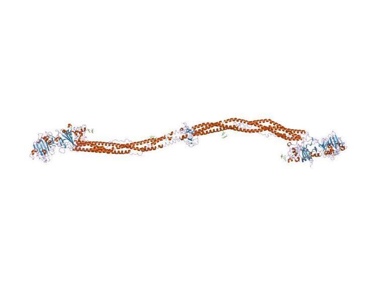 | ||
Fibrinogen (factor I) is a glycoprotein in vertebrates that helps in the formation of blood clots. It consists of a linear array of three nodules held together by a very thin thread which is estimated to have a diameter between 8 and 15 Angstrom (Å). The two end nodules are alike but the center one is slightly smaller. Measurements of shadow lengths indicate that nodule diameters are in the range 50 to 70 Å. The length of the dried molecule is 475 ± 25 Å.
Contents
The fibrinogen molecule is a soluble, large, and complex 340 kDa plasma glycoprotein, that is converted by thrombin into fibrin during blood clot formation. It has a rod-like shape with dimensions of 9 × 47.5 × 6 nm and it shows a negative net charge at physiological pH (IP at pH 5.2). Fibrinogen is synthesized in the liver by the hepatocytes. The concentration of fibrinogen in the blood plasma is 200–400 mg/dL (normally measured using the Clauss method).
During normal blood coagulation, a coagulation cascade activates the zymogen prothrombin by converting it into the serine protease thrombin. Thrombin then converts the soluble fibrinogen into insoluble fibrin strands. These strands are then cross-linked by factor XIII to form a blood clot. FXIIIa stabilizes fibrin further by incorporation of the fibrinolysis inhibitors alpha-2-antiplasmin and TAFI (thrombin activatable fibrinolysis inhibitor, procarboxypeptidase B), and binding to several adhesive proteins of various cells. Both the activation of factor XIII by thrombin and plasminogen activator (t-PA) are catalyzed by fibrin. Fibrin specifically binds the activated coagulation factors factor Xa and thrombin and entraps them in the network of fibers, thus functioning as a temporary inhibitor of these enzymes, which stay active and can be released during fibrinolysis. Research from 2011 has shown that fibrin plays a key role in the inflammatory response and development of rheumatoid arthritis.
Physiology
In its natural form, fibrinogen can form bridges between platelets, by binding to their GpIIb/IIIa surface membrane proteins; however, its major function is as the precursor to fibrin.
Fibrinogen, the principal protein of vertebrate blood clotting, is a hexamer, containing two sets of three different chains (α, β, and γ), linked to each other by disulfide bonds. The N-terminal sections of these three chains contain the cysteines that participate in the cross-linking of the chains. The C-terminal parts of the α, β and γ chains contain a domain of about 225 amino-acid residues, which can function as a molecular recognition unit. In fibrinogen as well as in angiopoietin, this domain is implicated in protein-protein interactions. In lectins, such as mammalian ficolins and invertebrate tachylectin 5A, the fibrinogen C-terminal domain binds carbohydrates. On the fibrinogen α and β chains, there is a small peptide sequence (called a fibrinopeptide). These small peptides are what prevent fibrinogen from spontaneously forming polymers with itself.
The conversion of fibrinogen to fibrin occurs in several steps. First, thrombin cleaves fibrinopeptide A and B located on the N-terminus of the fibrinogen alpha and beta chains respectively. The resulting fibrin monomers polymerize end to end to from protofibrils, which in turn associate laterally to form fibrin fibers. In a final step, the fibrin fibers associate to form the fibrin gel.
Fibrinogen deficiency
Congenital fibrinogen deficiency (afibrinogenemia) or disturbed function of fibrinogen has been described in a few cases.
It can lead to either bleeding or thromboembolic complications, or is clinically without pathological findings. More common are acquired deficiency stages that can be detected by laboratory tests in blood plasma or in whole blood by means of thrombelastometry. Acquired deficiency is found after hemodilution, blood losses and/or consumption such as in trauma patients, during some phases of disseminated intravascular coagulation (DIC), and also in sepsis. In patients with fibrinogen deficiency, the correction of bleeding is possible by infusion of fresh frozen plasma (FFP), cryoprecipitate (a fibrinogen-rich plasma fraction) or by fibrinogen concentrates. There is increasing evidence that correction of fibrinogen deficiency or fibrinogen polymerization disorders is very important in patients with bleeding.
Diagnostic use
Fibrinogen level can be measured in venous blood. Normal levels are about 1.5-3 g/L, depending on the method used. In typical circumstances, fibrinogen is measured in citrated plasma samples in the laboratory; however, the analysis of whole-blood samples by use of thromboelastometry (platelet function is inhibited with cytochalasin D) is also possible. Higher levels are, amongst others, associated with cardiovascular disease (>3.43 g/L). It may be elevated in any form of inflammation, as it is an acute-phase protein; for example, it is especially apparent in human gingival tissue during the initial phase of periodontal disease. Fibrinogen levels increase in pregnancy to an average of 4.5 g/l, compared to an average of 3 g/l in non-pregnant people.
It is used in veterinary medicine as an inflammatory marker: In horses, a level above the normal range of 1.0-4.0 g/L suggests some degree of systemic inflammatory response.
Low levels of fibrinogen can indicate a systemic activation of the clotting system, with consumption of clotting factors faster than synthesis. This excessive clotting factor consumption condition is known as disseminated intravascular coagulation or "DIC." DIC can be difficult to diagnose, but a strong clue is low fibrinogen levels in the setting of prolonged clotting times (PT or aPTT), in the context of acute critical illness such as sepsis or trauma. Besides low fibrinogen level, fibrin polymerization disorders that can be induced by several factors, including plasma expanders, can also lead to severe bleeding problems. Fibrin polymerization disorders can be detected by viscoelastic methods such as thrombelastometry.
A fibrinogen uptake test or fibrinogen scan is a test that was formerly used to detect deep vein thrombosis. In this method, radioactively labeled fibrinogen, typically with radioiodine, is given which is incorporated in the thrombus. The thrombus can then be detected by scintigraphy.