
 | ||
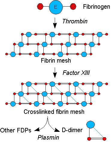
D-dimer (or D dimer) is a fibrin degradation product (or FDP), a small protein fragment present in the blood after a blood clot is degraded by fibrinolysis. It is so named because it contains two D fragments of the fibrin protein joined by a cross-link.
Contents
D-dimer concentration may be determined by a blood test to help diagnose thrombosis. Since its introduction in the 1990s, it has become an important test performed in patients with suspected thrombotic disorders. While a negative result practically rules out thrombosis, a positive result can indicate thrombosis but does not rule out other potential causes. Its main use, therefore, is to exclude thromboembolic disease where the probability is low. In addition, it is used in the diagnosis of the blood disorder disseminated intravascular coagulation.
Principles
Coagulation, the formation of a blood clot or thrombus, occurs when the proteins of the coagulation cascade are activated, either by contact with damaged blood vessel wall and exposure to collagen in the tissue space (extrinsic pathway) or by activation of factor VII by tissue activating factors (intrinsic pathway). Both pathways lead to the generation of thrombin, an enzyme that turns the soluble blood protein fibrinogen into fibrin, which aggregates into proteofibrils. Another thrombin-generated enzyme, factor XIII, then crosslinks the fibrin proteofibrils at the D fragment site, leading to the formation of an insoluble gel which serves as a scaffold for blood clot formation.
The circulating enzyme plasmin, the main enzyme of fibrinolysis, cleaves the fibrin gel in a number of places. The resultant fragments, "high molecular weight polymers", are digested several times more by plasmin to lead to intermediate and then to small polymers (fibrin degradation products or FDPs). The cross-link between two D fragments remains intact, however, and these are exposed on the surface when the fibrin fragments are sufficiently digested. The typical D-dimer containing fragment contains two D domains and one E domain of the original fibrinogen molecule.
D-dimers are not normally present in human blood plasma, except when the coagulation system has been activated, for instance because of the presence of thrombosis or disseminated intravascular coagulation. The D-dimer assay depends on the binding of a monoclonal antibody to a particular epitope on the D-dimer fragment. Several detection kits are commercially available; all of them rely on a different monoclonal antibody against D-dimer. For some of these, the area of the D-dimer to which the antibody binds is known. The binding of the antibody is then measured quantitatively by one of various laboratory methods.
Indications
D-dimer testing is of clinical use when there is a suspicion of deep venous thrombosis (DVT), pulmonary embolism (PE) or disseminated intravascular coagulation (DIC). It is under investigation in the diagnosis of aortic dissection.
For DVT and PE, there are possible various scoring systems that are used to determine the a priori clinical probability of these diseases; the best-known is the Wells score.
In some hospitals, they are measured by laboratories after a form is completed showing the probability score and only if the probability score is low or intermediate. This reduces the need for unnecessary tests in those who are high-probability. Performing the D-dimer test first can avoid a significant proportion of imaging tests and is less invasive. Since the D-dimer can exclude the need for imaging, specialty professional organizations recommend that physicians use D-dimer testing as an initial diagnostic.
Reference ranges
Following are reference ranges for D-dimer:
Thrombotic disease
Various kits have a 93-95% sensitivity (true positive rate). For hospitalized patients, one study found the specificity to be about 50% (true negative rate) in the diagnosis of thrombotic disease.
In interpretation of the d-dimer, for patients over age 50 a value of (patient's age) x 10 μg/l may be abnormal.
History
D-dimer was originally described in the 1970s, and found its diagnostic application in the 1990s.