
Entrez 3134 | Ensembl n/a | |
 | ||
Aliases HLA-F, CDA12, HLA-5.4, HLA-CDA12, HLA-F, major histocompatibility complex, class I, F External IDs MGI: 3779381 HomoloGene: 133121 GeneCards: HLA-F | ||
HLA class I histocompatibility antigen, alpha chain F is a protein that in humans is encoded by the HLA-F gene.
Contents
- HLA F
- Gene
- Protein
- Expression
- Intracellular expression
- Extracellular expression
- Expression during pregnancy
- Function
- Association with specialized ligands
- Maternal immunity tolerance
- Intermolecular communication
- Exogenous antigen cross presentation
- Ligand during inflammatory response
- Disease association
- References
HLA-F
The Major Histocompatibility Complex (MHC) is a group of cell surface proteins that in humans is also called the Human Leukocyte Antigen (HLA) complex. These proteins are encoded by a cluster of genes known as the HLA locus. The HLA locus occupies a ~ 3Mbp stretch that is located on the short arm of chromosome 6, specifically on 6p21.1-21.3. The MHC proteins are classified into three main categories, namely class I, II, and III. There are over 140 genes within the HLA locus and they are often called HLA genes. HLA-A, B, and C are the classical class I genes and HLA-E, F and G are the nonclassical class I genes. The protein encoded from the gene HLA-F was originally isolated from the human lymphoblastoid cell line 721.
Gene
The HLA-F gene is located on the short arm of chromosome 6, telomeric to the HLA-A locus. HLA-F has little allelic polymorphism and is highly conserved in other primates. HLA-F appears to be a recombinant between two multigene families, one that comprises conserved sequences found in all class I proteins (single transmembrane span) and another distinct family of genes with a conserved 3’ UTR. Many of these genes are highly transcribed and differentially expressed.
Protein
The HLA-F protein is a ~40-41 kDa molecule with conserved domains. Exon 7 is absent from the mRNA of HLA-F. The absence of this exon produces a modification in the cytoplasmic tail of the protein making it shorter relative to classical HLA class-I proteins. The cytoplasmic tail helps HLA-F exit the endoplasmic reticulum, and that function is primarily done by the amino acid valine found at the C-terminal end of the tail.
Expression
Classic HLA class I molecules interact with HLA-F through their heavy chain. However, HLA class I molecules only interact with HLA-F when they are in the form of an open conformer (free of peptide). Thus, HLA-F is expressed independently of bound peptide.
Intracellular expression
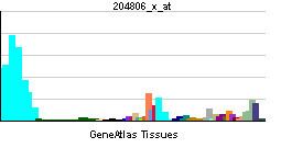
HLA-F is expressed intracellularly in peripheral blood lymphocytes (PBL), resting lymphocyte cells (B, T, NK, and monocytes), tonsils, spleen, thymus, bladder, brain, colon, kidney, liver, lymphoblast, T cell leukemia, choriocarcinoma, and carcinoma.
Extracellular expression
HLA-F is expressed on the cell surface of activated lymphocytes, HeLa cells, EBV-transformed lymphoblastoid cells, and in some activated monocyte cell lines. The surface expression of HLA-F coincides with the activated immune response since HLA-F is mostly found on the surface of stimulated T memory cells but not on circulating regulatory T cells.
Expression during pregnancy
HLA-F is expressed on the cells that surround the forming placenta (called extravillous trophoblasts), which are in direct contact with the maternal uterine cells. In these cells, HLA-F is expressed both intracellularly and on the surface.
Function
HLA-F belongs to the non-classical HLA class I heavy chain paralogues. Compared to classical HLA class I molecules, it exhibits very few polymorphisms. This class I molecule mainly exists as a heterodimer associated with the invariant light chain beta-2 microglobulin. The heavy chain is approximately 42 kDa and its gene contains 8 exons. Exon one encodes the leader peptide, exons 2 and 3 encode the alpha1 and alpha2 domains, the putative peptide binding sites, exon 4 encodes the alpha3 domain, exon 5 and 6 encode the transmembrane region and exons 7 and 8 the cytoplasmic tail. However, exons 7 and 8 (the cytoplasmic tail) are not translated due to an in-frame translation termination codon in exon 6.
HLA-F is currently the most enigmatic of the HLA molecules. Hence, its precise functions still remain to be resolved. Though, in contrast to other HLA molecules, it mainly resides intracellularly and rarely reaches the cell surface, e.g. upon activation of NK, B and T cells. Unlike classical HLA class I molecules, which possess ten highly conserved amino acids responsible for antigen recognition, HLA-F only has 5, suggesting a biological function different from peptide presentation. Upon immune cell activation, HLA-F binds free forms of HLA class I molecules and reaches the cell surface as heterodimer. In this way HLA-F stabilizes HLA class I molecules that haven't yet bound peptides, thereby acting as a chaperone and transporting the free HLA class I to, on, and from the cell surface.
Association with specialized ligands
HLA-F has been observed only in a subset of cell membranes, mostly B cells and activated lymphocytes. As a result, it has been suggested that its role involves association with specialized ligands that become available in the cell membrane of activated cells. For example, HLA-F can act as a peptide binding of ILT2 and ILT4. HLA-F can associate with TAP (transporter associated with antigen processing) and with the multimeric complex involved in peptide loading.
Maternal immunity tolerance
It has been observed that all three non-classical HLA class I proteins are expressed in placental trophoblasts in contact with maternal immune cells. This suggests that these proteins collaborate in the immune response and that HLA-F plays a fundamental role in both normal and maternal immune response. HLA-F is also expressed in decidual extravillous trophoblasts. During pregnancy, HLA-F interacts with T reg cells and extravillous trophoblasts mediating maternal tolerance to the fetus.
Intermolecular communication
During the interaction between HLA-F and the heavy chain (HC) of HLA class I molecules in activated lymphocytes, HLA-F plays a role as a chaperone, escorting HLA class I HC to the cell surface and stabilizing its expression in the absence of peptide. HLA-F binds most allelic forms of HLA class I open conformers, but it does not bind peptide complexes.
The expression patterns of HLA-F in T cells suggest that HLA-F is involved in the communication pathway between T reg and activated T cells, where HLA-F signals that the immune response has been activated. During this communication, either HLA-F invokes secretion of inhibitory cytokines by the regulatory T cells or it provides a simple inhibitory signal to the regulatory T cells, allowing a normal immune response to proceed.
Exogenous antigen cross-presentation
Viral proteins and other exogenous antigens decrease surface HLA-F expression because the exogenous proteins interact with HLA class I molecules at the same sites where HLA-F interacts, producing crosslinking. The exogenous proteins trigger an internal co-localization of both HLA-F and HLA class I molecules. Exogenous proteins with higher affinity will interact more readily with HLA class I molecules triggering a dissociation of HLA class I/HLA-F, thereby reducing the surface levels of HLA-F. HLA-F interacts with the open conformer (OC) of HLA class I and they function together in cross-presentation of exogenous antigen. Exogenous antigen binds to a structure on the surface of activated cells; this structure is composed of HLA class I open conformer and HLA-F; the peptide-binding point of contact is a specific HLA class I epitope on the exogenous antigen.
Ligand during inflammatory response
The complex HLA-F/HLA class I OC has two distinct roles that are central to the inflammatory response: first, it is a ligand for KIR receptors and can both activate and inhibit KIR; second, it is involved in cross-presentation of exogenous antigen.
The complex HLA-F/HLA class-I OC is a ligand for a subset of KIR (Killer-cell immunoglobulin-like receptor) receptors. Specifically, it was demonstrated that HLA-F interacts physically and functionally with three KIR receptors: KIR3DL2, KIR2DS4, and KIR3DS1, particularly during the inflammatory response. KIR directly interacts with both HLA-F and HLA class-I individually (i.e. no dimerization between HLA-F and HLA class-I is necessary).
Disease association
HLA-F has been linked to several diseases (Table). For cancer and tumors, HLA-F expression has been found to be enhanced in gastric adenocarcinoma, breast cancer, esophageal carcinoma, lung cancer, hepatocellular carcinoma, and neuroblastoma. HLA-F has also been associated with susceptibility to several diseases: hepatitis B, Systemic Lupus Erythematosus, and Type 1 diabetes (T1D).