
Entrez 2898 | Ensembl ENSG00000164418 | |
 | ||
Aliases GRIK2, EAA4, GLR6, GLUK6, GLUR6, GluK2, MRT6, glutamate ionotropic receptor kainate type subunit 2 External IDs MGI: 95815 HomoloGene: 40717 GeneCards: GRIK2 | ||
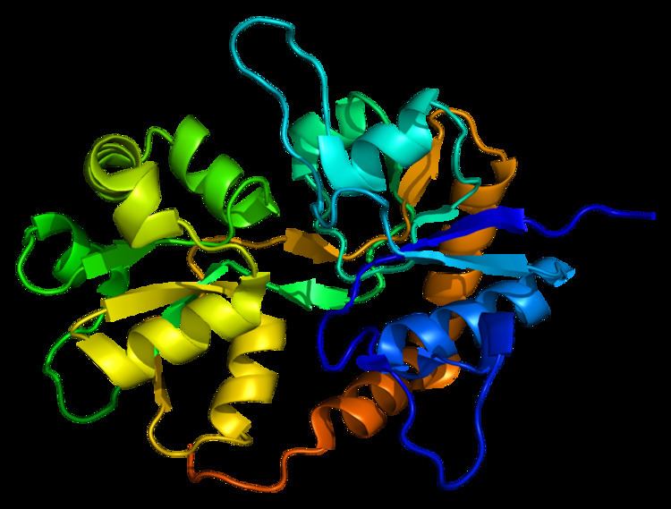
Glutamate receptor, ionotropic kainate 2 is a protein that in humans is encoded by the GRIK2 gene.
Contents
Function
This gene encodes a subunit of a kainate glutamate receptor. Glutamate receptors mediate the majority of excitatory neurotransmission in the brain. This receptor may have a role in synaptic plasticity and may be important for learning and memory. It also may be involved in the transmission of light information from the retina to the hypothalamus. The structure and function of the encoded protein is changed by RNA editing. Alternatively spliced transcript variants encoding distinct isoforms have been described for this gene.
Clinical significance
Homozygosity for a GRIK2 deletion-inversion mutation is associated with nonsyndromic autosomal recessive mental retardation.
Interactions
GRIK2 has been shown to interact with:
RNA Editing
Several ion channels and neurotransmitters receptors pre-mRNA as substrates for ADARs. This includes 5 subunits of the glutamate receptor ionotropic AMPA glutamate receptor subunits (Glur2, Glur3, Glur4) and kainate receptor subunits (Glur5, Glur6). Glutamate gated ion channels are made up of four subunits per channel with each subunit contributing to the pore loop structure. The pore loop structure is related to that found in K+ channels (e.g. human Kv1.1 channel). The human Kv1.1 channel pre mRNA is also subject to A to I RNA editing. The function of the glutamate receptors is in the mediation of fast neurotransmission to the brain. The diversity of the subunits is determined, as well as RNA splicing by RNA editing events of the individual subunits. This give rise to the necessarily high diversity of these receptors. Glur2 is a gene product of the pre- mRNA of the GRIK2 gene is subject to RNA editing.
Type
The type of RNA editing that occurs in the pre-mRNA of GluR-6 is Adenosine to Inosine ( A to I) editing.
A to I RNA editing is catalyzed by a family of adenosine deaminases acting on RNA (ADARs) that specifically recognize adenosines within double-stranded regions of pre-mRNAs and deaminate them to inosine. Inosines are recognised as guanosine by the cells translational machinery. There are three members of the ADAR family ADARs 1-3 with ADAR1 and ADAR2 being the only enzymatically active members. ADAR3 is thought to have a regulatory role in the brain. ADAR1 and ADAR2 are widely expressed in tissues while ADAR3 is restricted to the brain. The double-stranded regions of RNA are formed by base-pairing between residues in the close to region of the editing site with residues usually in a neighboring intron but can be an exonic sequence. The region that base pairs with the editing region is known as an Editing Complementary Sequence (ECS)
ADARs bind interact directly with the dsRNA substrate via their double-stranded RNA binding domains. If an editing site occurs within a coding sequence, the result could be a codon change. This can lead to translation of a protein isoform due to a change in its primary protein structure. Therefore, editing can also alter protein function. A to I editing occurs in a noncoding RNA sequences such as introns, untranslated regions (UTRs), LINEs, SINEs( especially Alu repeats) The function of A to I editing in these regions is thought to involve creation of splice sites and retention of RNAs in the nucleus amongst others.
Location
The pre-mRNA of GLUR-6 is edited at three positions at amino acid positions 567, 571, and 621. The Q/R position is so called as editing results in an codon change from a glutamine (Q) codon (CAG ) to an arginine (R) codon (CGG). This editing site is located in the " pore loop" of the second membrane domain (M2). Q/R editing site is also observed in glutamte receptor GluR-2 and GluR-5. The Q/R site of GluR-6 pre mRNA occurs in an asymmetrical loop of 3 exonic and four intronic nucleotides. The Q/R site is located in a homologous position in GluR-2 and in GluR-6.
GluR-6 is also edited at I/V and Y/C sites, which are found in the first membrane domain (M1). At the I/V site editing results in a codon change from (ATT) isoleucine (I) to (GTT)valine (V), while at the Y/C site the codon change is from (TAC) tyrosine(Y) to (TGC) cysteine (C).
The RNA fold programme characterised a putative double-stranded RNA(dsRNA) conformation around the Q/R site of the GluR-6 pre-mRNA. This sequence is necessary for editing at the site to occur. The possible editing complementary sequence was observed from transcript analysis to be 1.9 kb downstream from the editing site within intron 12. The ECS for the editing sites in M1 has yet to be identified but it is likely to occur at a considerable distance from the editing sites.
Regulation
Editing of the Q/R site in GluR-6 pre-mRNA has been demonstrated to be developmentally regulated in rats ranging from 0% in rat embryo to 80% at birth. This is different from AMPA receptor subunit GluR-B, which is edited nearly 100% and is not developmentally regulated. Significant amounts of both edited and nonedited forms of GluR-6 transcripts are found in adult brain. The receptor is 90% edited in all grey matter structures. In white matter of the brain the receptor is in edited form in just 10% of cases. Frequency increases from 0% in rat embryo to 85% in adult rat.
Structure
The primary of GluR-6 transcripts can be edited in up to three positions. Editing at each of the three positions affect the Ca2+ permeability of the channel
Function
Editing plays a role in the electrophysiology of the channel. Editing at the Q/R site has been deemed to be nonessential in GluR-6. It has been reported that unedited version of Glu-R6 functions in the regulation of synaptic plasticity. The edited version is thought to inhibit synaptic plasticity and reduce seizure susceptibility. Mice lacking the Q/R site are capable of long term potentiation and are more susceptible to kainate induced seizures. The number of seizures inversely correlates with the amount of rna editing. This correlates to the increase in human GluR-6 pre-mRNA editing during seizures. It is thought that editing maybe an adaptive mechanism.
Up to 8 different protein isoforms can occur as a result of different combinations of editing at the three sites. Editing at the Q/R site affects the calcium permeability of the receptor. The two other editing sites are less well defined (I/V,Y/C) but are also thought to be involved in regulation of calcium permeability.(59) Evidence suggests that if editing does not occur at I/V and Y/C sites then both edited and nonedited versions of the receptor demonstrate high calcium permeability. When both editing sites in TM1 are edited then the Q/R site edited version of the receptor is more permeable to calcium than the nonedited version at the Q/R site. The co assembly of these two isoforms generate receptor with reduced calcium permeability.
Rna editing of the Q/R site can effect inhibition of the channel by membrane fatty acids such as arachidonic acid and docosahexaenoic acid For Kainate receptors with only edited isforms, these are strongly inhibited by these fatty acids.However inclusion of just one nonedited subunit is enough to stop this inhibition.
Dysregulation
Kainate induced seizures in mice are used as a model of temporal lobe epilepsy in humans. Despite deficiency in editing at the Q/R site of GluR-6 in mice increasing seizure vulnerability, tissue analysis of human patients did not show reduced editing at this site.