
NeuroNames hier-429 Dorlands/Elsevier v_06/12853453 FMA 78454 | NeuroLex ID Third ventricle TA A14.1.08.410 | |
 | ||
Latin ventriculus tertius cerebri | ||
The third ventricle is one of four connected fluid-filled cavities comprising the ventricular system within the mammalian brain. It is a median cleft in the diencephalon between the two thalami, and is filled with cerebrospinal fluid (CSF).
Contents
It is in the midline, between the left and right lateral ventricles. Running through the third ventricle is the interthalamic adhesion, which contains thalamic neurons and fibers that may connect the two thalami.
Structure
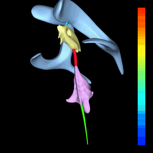
The third ventricle communicates with the lateral ventricles anteriorly by the interventricular foramina (of Monro). It also communicates with the fourth ventricle posteriorly by the cerebral aqueduct (of Sylvius).
Development
The third ventricle, like other parts of the ventricular system of the brain, develops from the central neural canal of the neural tube. Specifically, it originates from the portion of the tube that is present in the developing prosencephalon, and subsequently in the developing diencephalon.
Boundaries
The third ventricle is bounded by the thalamus and hypothalamus on both the left and right sides. The lamina terminalis forms the anterior wall. The floor is formed by hypothalamic structures. The roof is formed by the ependyma, lining the undersurface of the tela choroidea of the third ventricle.
Protrusions
There are two protrusions on the anterior aspect of the third ventricle:
Additionally, there are two protrusions on the posterior aspect, above the cerebral aqueduct:
In casts of the ventricular system, a small hole may be seen in the body of the third ventricle. This is formed where the two thalami are joined together at the interthalamic adhesion (not seen in all people).
Clinical significance
The floor of the third ventricle is formed by hypothalamic structures and this can be opened surgically between the mamillary bodies and the pituitary gland in a procedure called an endoscopic third ventriculostomy. An endoscopic third ventriculostomy can be performed in order to release extra fluid caused by hydrocephalus.
Several studies have found evidence of ventricular enlargement to be associated with major depression, particularly enlargement of the third ventricle. These observations are interpreted as indicating a loss of neural tissue in brain regions adjacent to the enlarged ventricle, leading to suggestions that cytokines and related mediators of neurodegeneration may play a role in giving rise to the disease.