
Latin fossa rhomboidea Dorlands/Elsevier f_14/12376599 FMA 78486 | NeuroNames hier-628 TA A14.1.05.702 | |
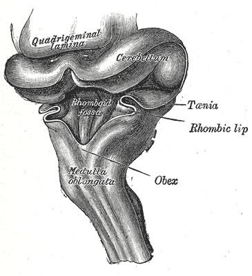 | ||
The rhomboid fossa is a rhombus-shaped depression that is the anterior part of the fourth ventricle. Its anterior wall, formed by the back of the pons and the medulla oblongata, constitutes the floor of the fourth ventricle.
Contents
It is covered by a thin layer of grey matter continuous with that of the spinal cord; superficial to this is a thin lamina of neuroglia which constitutes the ependyma of the ventricle and supports a layer of ciliated epithelium.
Parts
The fossa consists of three parts, superior, intermediate, and inferior.
The sulcus limitans forms the lateral boundary of the medial eminence.
Features
In the superior part of the rhomboid fossa it corresponds with the lateral limit of the fossa and presents a bluish-gray area, the locus coeruleus, which owes its color to an underlying patch of deeply pigmented nerve cells, termed the substantia ferruginea.
At the level of the facial colliculus the sulcus limitans widens into a flattened depression, the superior fovea, and in the inferior part of the fossa appears as a distinct dimple, the inferior fovea.
Lateral to the foveæ is a rounded elevation named the area acustica, which extends into the lateral recess and there forms a feebly marked swelling, the tuberculum acusticum.
Winding around the inferior peduncle and crossing the area acustica and the medial eminence are a number of white strands, the striæ medullares, which form a portion of the cochlear division of the acoustic nerve and disappear into the median sulcus.
Below the inferior fovea, and between the trigonum hypoglossi and the lower part of the area acustica is a triangular dark field, the vagal trigone, which corresponds to the sensory nucleus of the vagus and glossopharyngeal nerves.
The lower end of the vagal trigone is crossed by a narrow translucent ridge, the funiculus separans, and between this funiculus and the clava, is a small tongue-shaped area, the area postrema.
On section it is seen that the funiculus separans is formed by a strip of thickened ependyma, and the area postrema by loose, highly vascular, neuroglial tissue containing nerve cells of moderate size.