
Symbol Flavoprotein InterPro IPR003382 SUPERFAMILY 1e20 | Pfam PF02441 SCOP 1e20 Pfam structures | |
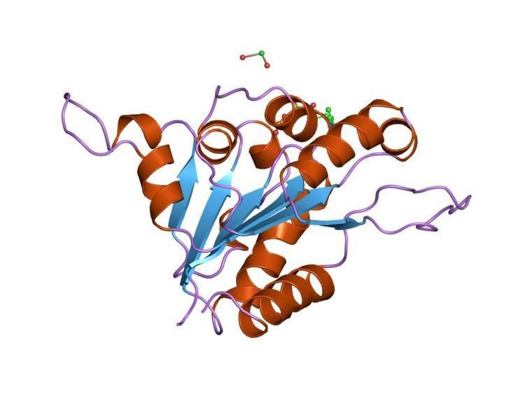 | ||
Flavoproteins are proteins that contain a nucleic acid derivative of riboflavin: the flavin adenine dinucleotide (FAD) or flavin mononucleotide (FMN).
Contents
Flavoproteins are involved in a wide array of biological processes, including, but by no means limited to, bioluminescence, removal of radicals contributing to oxidative stress, photosynthesis, DNA repair, and apoptosis. The spectroscopic properties of the flavin cofactor make it a natural reporter for changes occurring within the active site; this makes flavoproteins one of the most-studied enzyme families.
Flavoproteins have either an FMN or FAD molecule as a prosthetic group, this prosthetic group can be tightly bound or covalently linked. Only about 5-10% of flavoproteins have a covalently linked FAD, but these enzymes have stronger redox power. In some instances, FAD can provide structural support for active sites or provide stabilization of intermediates during catalysis. Based on the available structural data, the known FAD-binding sites can be divided into more than 200 different types.
There are 90 flavoproteins in the human genome; about 84% require FAD, and around 16% require FMN, whereas 5 proteins require both to be present. Flavoproteins are mainly located in the mitochondria because of their redox power. Of all flavoproteins, 90% perform redox reactions and the other 10% are transferases, lyases, isomerases, ligases.
Discovery
The first mention of a flavoprotein in the scientific literature dates back to 1879, when the work on the composition of cow’s milk resulted in the isolation of a bright-yellow pigment, that we now know as flavin, but termed lactochrome at the time. By the early 1930s, this same pigment had been isolated from a range of sources, and recognised as a component of the vitamin B complex. Its structure was determined almost simultaneously by two groups in 1934, and given the name riboflavin, derived from the ribityl side chain and yellow colour of the conjugated ring system.
The first evidence for the requirement of flavin as an enzyme cofactor came in 1935. Hugo Theorell and coworkers showed that a bright-yellow-coloured yeast protein, identified previously as essential for cellular respiration, could be separated into apoprotein and a bright-yellow pigment. Neither apoprotein nor pigment alone could catalyse the oxidation of NADH, but mixing of the two restored the enzyme activity. However, replacing the isolated pigment with riboflavin did not restore enzyme activity, despite their being indistinguishable under spectroscopy. This led to the discovery that the protein studied required not riboflavin but flavin mononucleotide to be catalytically active
Similar experiments with D-amino acid oxidase led to the identification of flavin adenine dinucleotide (FAD) as a second form of flavin utilised by enzymes.
Examples
The flavoprotein family contains a diverse range of enzymes, including: