The femoral nerve is a nerve in the thigh that supplies skin on the upper thigh and inner leg, and the muscles that extend the knee.
The femoral nerve is the largest branch of the lumbar plexus, and arises from the dorsal divisions of the ventral rami of the second, third, and fourth lumbar nerves (L2-L4).
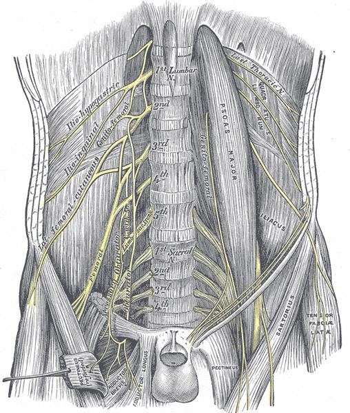
It descends through the fibers of the psoas major muscle, emerging from the muscle at the lower part of its lateral border, and passes down between it and the iliacus muscle, behind the iliac fascia; it then runs beneath the inguinal ligament, into the thigh, and splits into an anterior and a posterior division. Under the inguinal ligament, it is separated from the femoral artery by a portion of the psoas major.
The muscles innervated by the femoral nerve extend the knee. The nerve is also responsible for sensation over the front and inner sides of the thigh, shin, and arch of the foot.
Within the abdomen the femoral nerve gives off small branches to the iliacus muscle, and a branch which is distributed on the upper part of the femoral artery; the latter branch may arise in the thigh.
AnteriorIn the thigh, the anterior division of the femoral nerve gives off anterior cutaneous and muscular branches.
Anterior cutaneous branches: The anterior cutaneous branches comprise the following nerves: intermediate femoral cutaneous nerve and medial femoral cutaneous nerve (Note the lateral femoral cutaneous nerve is a branch from the lumbar plexus.)Muscular branches: The nerve to the Pectineus arises immediately below the inguinal ligament, and passes behind the femoral sheath to enter the anterior surface of the muscle; it is often duplicated. The nerve to the Sartorius arises in common with the intermediate cutaneous.PosteriorThe femoral nerve has two groups of posterior branches, muscular (to the four parts of the Quadriceps femoris), and articular, to the knee.
Muscular branches to the quadriceps include:
The branch to the Rectus femoris enters the upper part of the inner surface of the muscle, and supplies a filament to the hip.A large branch to the Vastus lateralis accompanies the descending branch of the lateral femoral circumflex artery to the lower part of the muscle. It gives off an articular filament to the knee.The branch to the Vastus medialis descends lateral to the femoral vessels in company with the saphenous nerve. It enters the muscle about its middle, and gives off a filament, which can usually be traced downward, on the surface of the muscle, to the knee.The branches to the Vastus intermedius, two or three in number, enter the anterior surface of the muscle about the middle of the thigh; a filament from one of these descends through the muscle to the Articularis genus and the knee-joint. The articular branch to the hip-joint is derived from the nerve to the Rectus femoris.There are three articular branches:
A long slender filament, derived from the nerve to the Vastus lateralis; it penetrates the capsule of the joint on its anterior aspect.A branch derived from the nerve to the Vastus medialis, can usually be traced downward on the surface of this muscle to near the joint; it then penetrates the muscular fibers, and accompanies the articular branch of the highest genicular artery, pierces the medial side of the articular capsule, and supplies the synovial membrane.A third branch is derived from the nerve to the Vastus intermedius.