
Antagonist Gluteus maximus | ||
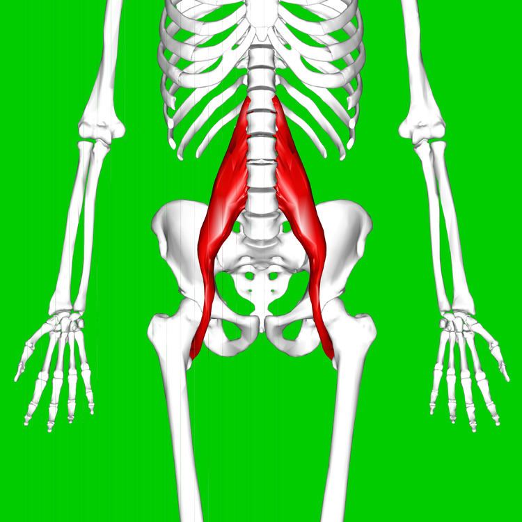 | ||
Origin Transverse processes of T12-L5 and the lateral aspects of the discs between them Actions Flexion in the hip joint | ||
The psoas major (/ˈsoʊ.əs/ or /ˈsoʊ.æs/) (from Greek: ψόας - psóās : 'of the loins', genitive singular form of ψόα - psóa 'the loins') is a long fusiform muscle located on the side of the lumbar region of the vertebral column and brim of the lesser pelvis. It joins the iliacus muscle to form the iliopsoas.
Contents
Structure
The psoas major is divided into a superficial and deep part. The deep part originates from the transverse processes of lumbar vertebrae I-V. The superficial part originates from the lateral surfaces of the last thoracic vertebra, lumbar vertebrae I-IV, and from neighboring intervertebral discs. The lumbar plexus lies between the two layers.
The iliacus and psoas major form the iliopsoas, which is surrounded by the iliac fascia. The iliopsoas runs across the iliopubic eminence through the muscular lacuna to its insertion on the lesser trochanter of the femur. The iliopectineal bursa separates the tendon of the iliopsoas muscle from the external surface of the hip joint capsule at the level of the iliopubic eminence. The iliac subtendinous bursa lies between the lesser trochanter and the attachment of the iliopsoas.
Innervation
Innervation of the psoas major is through the anterior rami of L1 to L3 nerves.
Variation
In less than 50 percent of human subjects, the psoas major is accompanied by the psoas minor.
In animals
In mice, it is mostly a fast-twitching, type II muscle, while in human it combines slow and fast-twitching fibers.
This muscle is equivalent to the tenderloin.
Function
The psoas major joins the upper body and the lower body, the axial to the appendicular skeleton, the inside to the outside, and the back to the front. As part of the iliopsoas, psoas major contributes to flexion in the hip joint. On the lumbar spine, unilateral contraction bends the trunk laterally, while bilateral contraction raises the trunk from its supine position.
It forms part of a group of muscles called the hip flexors, whose action is primarily to lift the upper leg towards the body when the body is fixed or to pull the body towards the leg when the leg is fixed.
For example, when doing a sit-up that brings the torso (including the lower back) away from the ground and towards the front of the leg, the hip flexors (including the iliopsoas) will flex the spine upon the pelvis.
Owing to the frontal attachment on the vertebrae, rotation of the spine will stretch the psoas.
Clinical significance
Tightness of the psoas can result in spasms or lower back pain by compressing the lumbar discs. A hypertonic and inflamed psoas can lead to irritation and entrapment of the iliolinguinal and the iliohypogastric nerves, resulting in a sensation of heat or water running down the front of the thigh.
Psoas can be palpated with active flexion of the hip. A positive psoas contracture test and pain with palpation reported by the patient indicate clinical significance. Care should be taken around the abdominal organs, especially the colon when palpating deeply.
The appearance of a protruding belly can visually indicate a hypertonic psoas, which pulls the spine forward while pushing the abdominal contents outward.
Racial variation
Autopsy data show this muscle is thicker in those of African descent than in Caucasians, and that the presence of the psoas minor is also racially variant.