
Latin Arteria femoralis | Vein Femoral vein MeSH A07.231.114.351 | |
 | ||
Branches Superficial epigastric artery, superficial iliac circumflex, superficial external pudendal, deep external pudendal, deep femoral artery, continues as popliteal artery Supplies Anterior compartment of thigh | ||
The femoral artery (Latin: arteria femoralis) is a large artery in the thigh and the main arterial supply to the lower limb. It enters the thigh from behind the inguinal ligament as the common femoral artery, a continuation of the external iliac artery. Here, it lies midway between the anterior superior iliac spine and the symphysis pubis. The common femoral artery gives off the profunda femoris artery and becomes the superficial femoral artery to descend along the anteromedial part of the thigh in the femoral triangle. It enters and passes through the adductor (subsartorial) canal, and becomes the popliteal artery as it passes through an opening in adductor magnus near the junction of the middle and distal thirds of the thigh.
Contents
Structure
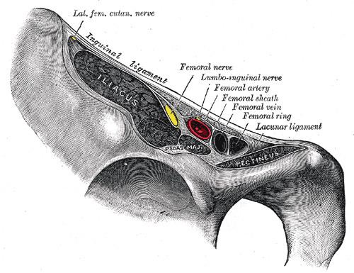
Its first three or four centimetres are enclosed, with the femoral vein, in the femoral sheath. The relations of the femoral artery are as follows:
The femoral artery gives off several branches in the thigh which include;
In clinical parlance, the part of the femoral artery proximal to the origin of profunda femoris is often termed the common femoral artery, while that distal to the origin of the profunda is termed the superficial femoral artery.
Pulse
As the femoral artery can often be palpated through the skin, it is often used as a catheter access artery. From it, wires and catheters can be directed anywhere in the arterial system for intervention or diagnostics, including the heart, brain, kidneys, arms and legs. The direction of the needle in the femoral artery can be against blood flow (retro-grade), for intervention and diagnostic towards the heart and opposite leg, or with the flow (ante-grade or ipsi-lateral) for diagnostics and intervention on the same leg. Access in either the left or right femoral artery is possible and depends on the type of intervention or diagnostic.
The site for optimally palpating the femoral pulse is in the inner thigh, at the mid-inguinal point, halfway between the pubic symphysis and anterior superior iliac spine. Presence of a femoral pulse has been estimated to indicate a systolic blood pressure of more than 50 mmHg, as given by the 50% percentile.
The femoral artery can be used to draw arterial blood when the blood pressure is so low that the radial or brachial arteries cannot be located.
Peripheral arterial disease
The femoral artery is susceptible to peripheral arterial disease. When it is blocked through atherosclerosis, percutaneous intervention with access from the opposite femoral may be needed. Endarterectomy, a surgical cut down and removal of the plaque of the femoral artery is also common. If the femoral artery has to be ligated surgically to treat a popliteal aneurysm, blood can still reach the popliteal artery distal to the ligation via the genicular anastomosis. However, if flow in the femoral artery of a normal leg is suddenly disrupted, blood flow distally is rarely sufficient. The reason for this is the fact that the genicular anastomosis is only present in a minority of individuals and is always undeveloped when disease in the femoral artery is absent.
History
Textbook illustrations of the genicular anastomosis, such as that shown in the sidebox, all appear to have been derived from the idealized image first produced by Gray's Anatomy in 1910. Neither the 1910 illustration, nor any subsequent version, was made of an anatomical dissection but rather from the writings of John Hunter (surgeon) and Astley Cooper which described the genicular anastomosis many years after ligation of the femoral artery for popliteal aneurysm. The genicular anastomosis has not been demonstrated even with modern imaging techniques such as X-ray computed tomography or angiography.