
 | ||
The ErbB family of proteins contains four receptor tyrosine kinases, structurally related to the epidermal growth factor receptor (EGFR), its first discovered member. In humans, the family includes Her1 (EGFR, ErbB1), Her2 (Neu, ErbB2), Her3 (ErbB3), and Her4 (ErbB4). The gene symbol, ErbB, is derived from the name of a viral oncogene to which these receptors are homologous: erythroblastic leukemia viral oncogene. Insufficient ErbB signaling in humans is associated with the development of neurodegenerative diseases, such as multiple sclerosis and Alzheimer's Disease, while excessive ErbB signaling is associated with the development of a wide variety of types of solid tumor.
Contents
In mice, loss of signaling by any member of the ErbB family results in embryonic lethality with defects in organs including the lungs, skin, heart, and brain. Excessive ErbB signaling is associated with the development of a wide variety of types of solid tumor. ErbB-1 and ErbB-2 are found in many human cancers, and their excessive signaling may be critical factors in the development and malignancy of these tumors.
Family members
The ErbB protein family consists of 4 members
v-ErbBs are homologous to EGFR, but lack sequences within the ligand binding ectodomain.
Structure
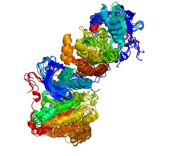
ErbB receptors are made up of an extracellular region or ectodomain that contains approximately 620 amino acids, a single transmembrane-spanning region, and a cytoplasmic tyrosine kinase domain. The extracellular region of each family member is made up of four subdomains, L1, CR1, L2, and CR2, where "L" signifies a leucine-rich repeat domain and "CR" a cysteine-rich region. These subdomains are shown in blue (L1), green (CR1), yellow (L2), and red (CR2) in the figure below. These subdomains are also referred to as domains I-IV, respectively.
The figure below shows the tridimensional structure of the ErbB family proteins, using the pdb files 1NQL (ErbB-1), 1S78 (ErbB-2), 1M6B (ErbB-3) and 2AHX (ErbB-4):
Kinase activation
The four members of the ErbB protein family are capable of forming homodimers, heterodimers, and possibly higher-order oligomers upon activation by a subset of potential growth factor ligands. There are 11 growth factors that activate ErbB receptors.
The ability ('+') or inability ('-') of each growth factor to activate each of the ErbB receptors is shown in the table below:
When not bound to a ligand, the extracellular regions of ErbB-1, -3, and -4 are found in a tethered conformation in which a 10-amino-acid-long dimerisation arm is unable to mediate monomer-monomer interactions. In contrast, in ligand-bound ErbB-1 and unliganded ErbB-2, the dimerisation arm becomes untethered and exposed at the receptor surface, making monomer-monomer interactions and dimerisation possible. The consequence of ectodomain dimerisation is the positioning of two cytoplasmic domains such that transphosphorylation of specific tyrosine, serine, and threonine amino acids can occur within the cytoplasmic domain of each ErbB. At least 10 specific tyrosines, 7 serines, and 2 threonines have been identified within the cytoplamic domain of ErbB-1, that may become phosphorylated and in some cases de-phosphorylated (e.g., Tyr 992) upon receptor dimerisation. Although a number of potential phosphorylation sites exist, upon dimerisation only one or much more rarely two of these sites are phosphorylated at any one time.
Role in cancer
Phosphorylated tyrosine residues act as binding sites for intracellular signal activators such as Ras. The Ras-Raf-MAPK pathway is a major signalling route for the ErbB family, as is the PI3-K/AKT pathway, both of which lead to increased cell proliferation and inhibition of apoptosis.
ErbB-1 is overexpressed in many cancers. Drugs such as panitumumab, cetuximab, gefitinib, erlotinib, afatinib are used to inhibit it. It has recently been shown that acquired resistance to cetuximab and gefitinib can be linked to hyperactivity of ErbB-3. This is linked to an acquired overexpression of c-MET, which phosphorylates ErbB-3, which in turn activates the AKT pathway.
ErbB-2 (HER-2) is often overexpressed in breast cancer, and is targeted by the drug trastuzumab (Herceptin). Only one-third of the women respond to trastuzumab. While the mechanism of resistance has not yet been elucidated, it has been shown that patients with ER+/HER2+ compared with ER-/HER2+ breast cancers may actually benefit more from drugs that inhibit the PI3K/AKT molecular pathway.