
EC number 4.2.1.11 ExPASy NiceZyme view | CAS number 9014-08-8 | |
 | ||
Enolase, also known as phosphopyruvate hydratase, is a metalloenzyme responsible for the catalysis of the conversion of 2-phosphoglycerate (2-PG) to phosphoenolpyruvate (PEP), the ninth and penultimate step of glycolysis. The chemical reaction catalyzed by enolase is:
Contents
2-phospho-D-glycerateEnolase belongs to the family of lyases, specifically the hydro-lyases, which cleave carbon-oxygen bonds. The systematic name of this enzyme is 2-phospho-D-glycerate hydro-lyase (phosphoenolpyruvate-forming).
The reaction is reversible, depending on environmental concentrations of substrates. The optimum pH for the human enzyme is 6.5. Enolase is present in all tissues and organisms capable of glycolysis or fermentation. The enzyme was discovered by Lohmann and Meyerhof in 1934, and has since been isolated from a variety of sources including human muscle and erythrocytes. In humans, deficiency of ENO1 is linked to hereditary haemolytic anemia while ENO3 deficiency is linked to glycogen storage disease XIII.
Isozymes
In humans there are three subunits of enolase, α, β, and γ, each encoded by a separate gene that can combine to form five different isoenzymes: αα, αβ, αγ, ββ, and γγ. Three of these isoenzymes (all homodimers) are more commonly found in adult human cells than the others:
When present in the same cell, different isozymes readily form heterodimers.
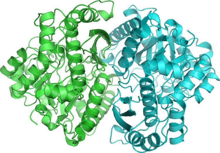
Structure
Enolase is a member of the large enolase superfamily. It has a molecular weight of 82,000-100,000 Daltons depending on the isoform. In human alpha enolase, the two subunits are antiparallel in orientation so that Glu20 of one subunit forms an ionic bond with Arg414 of the other subunit. Each subunit has two distinct domains. The smaller N-terminal domain consists of three α-helices and four β-sheets. The larger C-terminal domain starts with two β-sheets followed by two α-helices and ends with a barrel composed of alternating β-sheets and α-helices arranged so that the β-beta sheets are surrounded by the α-helices. The enzyme’s compact, globular structure results from significant hydrophobic interactions between these two domains.
Enolase is a highly conserved enzyme with five active-site residues being especially important for activity. When compared to wild-type enolase, a mutant enolase that differs at either the Glu168, Glu211, Lys345, or Lys396 residue has an activity level that is cut by a factor of 105. Also, changes affecting His159 leave the mutant with only 0.01% of its catalytic activity. An integral part of enolase are two Mg2+ cofactors in the active site, which serve to stabilize negative charges in the substrate.
Recently, moonlighting functions of several enolases, such as interaction with plasminogen, have brought interest to the enzymes' catalytic loops and their structural diversity.
Mechanism
Using isotopic probes, the overall mechanism for converting 2-PG to PEP is proposed to be an E1cb elimination reaction involving a carbanion intermediate. The following detailed mechanism is based on studies of crystal structure and kinetics. When the substrate, 2-phosphoglycerate, binds to α-enolase, its carboxyl group coordinates with two magnesium ion cofactors in the active site. This stabilizes the negative charge on the deprotonated oxygen while increasing the acidity of the alpha hydrogen. Enolase’s Lys345 deprotonates the alpha hydrogen, and the resulting negative charge is stabilized by resonance to the carboxylate oxygen and by the magnesium ion cofactors. Following the creation of the carbanion intermediate, the hydroxide on C3 is eliminated as water with the help of Glu211, and PEP is formed.
Additionally, conformational changes occur within the enzyme that aid catalysis. In human α-enolase, the substrate is rotated into position upon binding to the enzyme due to interactions with the two catalytic magnesium ions, Gln167, and Lys396. Movements of loops Ser36 to His43, Ser158 to Gly162, and Asp255 to Asn256 allow Ser39 to coordinate with Mg2+ and close off the active site. In addition to coordination with the catalytic magnesium ions, the pKa of the substrate’s alpha hydrogen is also lowered due to protonation of the phosphoryl group by His159 and its proximity to Arg374. Arg374 also causes Lys345 in the active site to become deprotonated, which primes Lys345 for its role in the mechanism.
Diagnostic uses
In recent medical experiments, enolase concentrations have been sampled in an attempt to diagnose certain conditions and their severity. For example, higher concentrations of enolase in cerebrospinal fluid more strongly correlated to low-grade astrocytoma than did other enzymes tested (aldolase, pyruvate kinase, creatine kinase, and lactate dehydrogenase). The same study showed that the fastest rate of tumor growth occurred in patients with the highest levels of CSF enolase. Increased levels of enolase have also been identified in patients who have suffered a recent myocardial infarction or cerebrovascular accident. It has been inferred that levels of CSF neuron-specific enolase, serum NSE, and creatine kinase (type BB) are indicative in the prognostic assessment of cardiac arrest victims. Other studies have focused on the prognostic value of NSE values in cerebrovascular accident victims.
Autoantibodies to alpha-enolase are associated with the rare syndrome called Hashimoto's encephalopathy.
Inhibitors
Small-molecule inhibitors of enolase have been synthesized as chemical probes (substrate-analogues) of the catalytic mechanism of the enzyme and more recently, have been investigated as potential treatments for cancer and infectious diseases. Most inhibitors have metal chelating properties and bind to enzyme by interactions with the structural Magnesium Atom Mg(A). The most potent of these is phosphonoacetohydroxamate, which in its unprotonated form has pM affinity for the enzyme. It has structural similarity to the presumed catalytic intermediate, between PEP and 2-PG. Attempts have been made to use this inhibitor as an anti-trypanosome drug, and more recently, as an anti-cancer agent, specifically, in glioblastoma that are enolase-deficient due to homozygous deletion of the ENO1 gene as part of the 1p36 tumor suppressor locus (synthetic lethality). A natural product phosphonate antibiotic, SF2312 (CAS 107729-45-3), which is active against gram positive and negative bacteria especially under anaerobic conditions, is a high potency inhibitor of Enolase 4zcw that binds in manner similar to phoshphonoacetohydroxamate 4za0. An allosteric binder, ENOblock was initially described as an inhibitor of Enolase, but subsequently shown not to actually inhibit the enzyme, but rather, interfere with the Enolase in vitro enzymatic assay.
Fluoride is a known competitor of enolase’s substrate 2-PG. The fluoride is part of a complex with magnesium and phosphate, which binds in the active site instead of 2-PG. As such, drinking fluoridated water provides fluoride at a level that inhibits oral bacteria enolase activity. Disruption of the bacteria’s glycolytic pathway - and, thus, its normal metabolic functioning - prevents dental caries from forming.