
ICD-9-CM 191 OMIM 137800 | ICD-10 C71 ICD-O M9440/3 | |
 | ||
Synonyms glioblastoma multiforme, grade IV astrocytoma | ||
Glioblastoma, also known as glioblastoma multiforme (GBM), is the most aggressive cancer that begins within the brain. Signs and symptoms of glioblastoma are initially non-specific. They may include headaches, personality changes, nausea, and symptoms similar to those of a stroke. Worsening of symptoms is often rapid. This can progress to unconsciousness.
Contents
- Signs and symptoms
- Risk factors
- Pathogenesis
- Molecular alterations
- Glioblastoma stem like cells
- Metabolism
- Ion channels
- Diagnosis
- Treatment
- Symptomatic therapy
- Palliative therapy
- Surgery
- Radiotherapy
- Chemotherapy
- Other modalities
- Prognosis
- Epidemiology
- History
- Research
- MicroRNA
- Immunotherapy
- Gene therapy
- Intranasal drug delivery
- References
The cause of most cases is unclear. Uncommon risk factors include genetic disorders such as neurofibromatosis and Li Fraumeni syndrome and previous radiation therapy. Glioblastomas represent 15% of brain tumors. They can either start from normal brain cells or develop from an already existing low-grade astrocytoma. The diagnosis is typically made by a combination of CT scan, MRI scan, and tissue biopsy.
There is no clear way to prevent the disease. Typically treatment involves surgery after which chemotherapy and radiation therapy are used. The medication temozolomide is frequently used as part of chemotherapy. High dose steroids may be used to help reduce swelling and decrease symptoms. It is unclear whether trying to remove all or simply most of the cancer is better.
Despite maximum treatment, the cancer usually recurs. The most common length of survival following diagnosis is 12 to 15 months with less than 3% to 5% of people surviving longer than five years. Without treatment survival is typically 3 months. It is the most common cancer that begins within the brain and the second most common brain tumor after meningioma. About 3 per 100,000 people develop the disease a year. It most often begins around 64 years of age and occurs more commonly in males than females. Immunotherapy is being studied in glioblastoma with promising results.
Signs and symptoms
Although common symptoms of the disease include seizure, nausea and vomiting, headache, memory loss, and hemiparesis, the single most prevalent symptom is a progressive memory, personality, or neurological deficit due to temporal and frontal lobe involvement. The kind of symptoms produced depends more on the location of the tumor than on its pathological properties. The tumor can start producing symptoms quickly, but occasionally is an asymptomatic condition until it reaches an enormous size.
Risk factors
For unknown reasons, GBM occurs more commonly in males. Most glioblastoma tumors appear to be sporadic, without any genetic predisposition. No links have been found between glioblastoma and smoking, consumption of cured meat, or electromagnetic fields. Alcohol consumption may be a possible risk factor. Glioblastoma has been associated with the viruses SV40, HHV-6, and cytomegalovirus. There also appears to be a small link between ionizing radiation and glioblastoma. A 2006 analysis links brain cancer to lead exposure in the work-place. There is an association of brain tumor incidence and malaria, suggesting that the anopheles mosquito, the carrier of malaria, might transmit a virus or other agent that could cause glioblastoma or that the immunosuppression associated with malaria could enhance viral replication. Also HHV-6 reactivates in response to hypersensitivity reactions from drugs and environmental chemicals.
Other risk factors include:
Pathogenesis
Glioblastoma multiforme tumors are characterized by the presence of small areas of necrotizing tissue that are surrounded by anaplastic cells. This characteristic, as well as the presence of hyperplastic blood vessels, differentiates the tumor from Grade 3 astrocytomas, which do not have these features.
GBMs usually form in the cerebral white matter, grow quickly, and can become very large before producing symptoms. Less than 10% form more slowly following degeneration of low-grade astrocytoma or anaplastic astrocytoma. These are called secondary GBMs and are more common in younger patients (mean age 45 versus 62 years). The tumor may extend into the meninges or ventricular wall, leading to high protein content in the cerebrospinal fluid (CSF) (> 100 mg/dL), as well as an occasional pleocytosis of 10 to 100 cells, mostly lymphocytes. Malignant cells carried in the CSF may spread (rarely) to the spinal cord or cause meningeal gliomatosis. However, metastasis of GBM beyond the central nervous system is extremely unusual. About 50% of GBMs occupy more than one lobe of a hemisphere or are bilateral. Tumors of this type usually arise from the cerebrum and may exhibit the classic infiltration across the corpus callosum, producing a butterfly (bilateral) glioma.
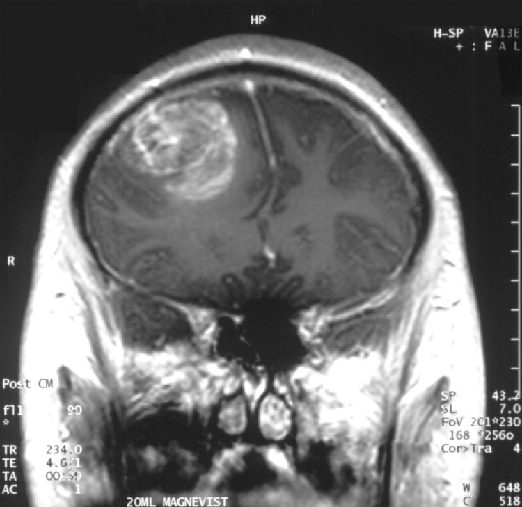
The tumor may take on a variety of appearances, depending on the amount of hemorrhage, necrosis, or its age. A CT scan will usually show an inhomogeneous mass with a hypodense center and a variable ring of enhancement surrounded by edema. Mass effect from the tumor and edema may compress the ventricles and cause hydrocephalus.
Molecular alterations
Four subtypes of glioblastoma have been identified:
Many other genetic alterations have been described in glioblastoma, and the majority of them are clustered in two pathways, the RB and the PI3K/AKT. Glioblastomas have alterations in 68-78% and 88% of these pathways, respectively.
Another important alteration is methylation of MGMT, a "suicide" DNA repair enzyme. Methylation is described to impair DNA transcription and therefore, expression of the MGMT enzyme. Since an MGMT enzyme can only repair one DNA alkylation due to its suicide repair mechanism, reverse capacity is low and methylation of the MGMT gene promoter greatly affects DNA-repair capacity. Indeed, MGMT methylation is associated with an improved response to treatment with DNA-damaging chemotherapeutics, such as temozolomide.
Glioblastoma stem-like cells
Cancer cells with stem cell-like properties have been found in glioblastomas (this may be a cause of their resistance to conventional treatments, and high recurrence rate). These so-called glioblastoma stem-like cells reside in a niche around arterioles, which protects these cells against therapy by maintaining a relatively hypoxic environment. A biomarker for cells in glioblastomas that exhibit cancer stem cell properties, the transcription factor Hes3, has been shown to regulate their number when placed in culture.
Metabolism
The IDH1 gene encodes for the enzyme isocitrate dehydrogenase 1 and is frequently mutated in glioblastoma (primary GBM: 5%, secondary GBM >80%). By producing very high concentrations of the "oncometabolite" D-2-hydroxyglutarate and dysregulating the function of the wild-type IDH1-enzyme it induces profound changes to the metabolism of IDH1-mutated glioblastoma, compared with IDH1 wild-type glioblastoma or healthy astrocytes. Among others, it increases the glioblastoma cells' dependence on glutamine or glutamate as an energy source. It has been hypothesized that IDH1-mutated glioblastoma are in a very high demand for glutamate and use this amino acid and neurotransmitter as a chemotactic signal. Since healthy astrocytes excrete glutamate, IDH1-mutated glioblastoma cells do not favor dense tumor structures but instead migrate, invade and disperse into healthy parts of the brain where glutamate concentrations are higher. This may explain the invasive behaviour of these IDH1-mutated glioblastoma.
Ion channels
Furthermore, glioblastoma multiforme exhibits numerous alterations in genes that encode for ion channels, including upregulation of gBK potassium channels and ClC-3 chloride channels. It has been hypothesized that by upregulating these ion channels, glioblastoma tumor cells can facilitate increased ion movement over the cell membrane, thereby increasing H2O movement through osmosis, which aids glioblastoma cells in changing cellular volume very rapidly. This is helpful in their extremely aggressive invasive behavior, because quick adaptations in cellular volume can facilitate movement through the sinuous extracellular matrix of the brain.
Diagnosis
When viewed with MRI, glioblastomas often appear as ring-enhancing lesions. The appearance is not specific, however, as other lesions such as abscess, metastasis, tumefactive multiple sclerosis, and other entities may have a similar appearance. Definitive diagnosis of a suspected GBM on CT or MRI requires a stereotactic biopsy or a craniotomy with tumor resection and pathologic confirmation. Because the tumor grade is based upon the most malignant portion of the tumor, biopsy or subtotal tumor resection can result in undergrading of the lesion. Imaging of tumor blood flow using perfusion MRI and measuring tumor metabolite concentration with MR spectroscopy may add value to standard MRI in select cases by showing increased relative cerebral blood volume and increased choline peak respectively, but pathology remains the gold standard for diagnosis and molecular characterization.
It is important to distinguish primary glioblastoma from secondary glioblastoma. These tumors occur spontaneously (de novo) or have progressed from a lower-grade glioma, respectively. Primary glioblastomas have a worse prognosis, different tumor biology and may have a different response to therapy, which makes this a critical evaluation to determine patient prognosis and therapy. Over 80% of secondary glioblastoma carries a mutation in IDH1, whereas this mutation is rare in primary glioblastoma (5-10%). Thus, IDH1 mutations are a useful tool to distinguish primary and secondary glioblastomas since histopathologically they are very similar and the distinction without molecular biomarkers is unreliable.
Treatment
It is very difficult to treat glioblastoma due to several complicating factors:
Treatment of primary brain tumors and brain metastases consists of both symptomatic and palliative therapies.
Symptomatic therapy
Supportive treatment focuses on relieving symptoms and improving the patient’s neurologic function. The primary supportive agents are anticonvulsants and corticosteroids.
Palliative therapy
Palliative treatment usually is conducted to improve quality of life and to achieve a longer survival time. It includes surgery, radiation therapy, and chemotherapy. A maximally feasible resection with maximal tumor-free margins is usually performed along with external beam radiation and chemotherapy. Gross total resection of tumor is associated with a better prognosis.
Surgery
Surgery is the first stage of treatment of glioblastoma. An average GBM tumor contains 1011 cells, which is on average reduced to 109 cells after surgery (a reduction of 99%). Benefits of surgery include resection for a pathological diagnosis, alleviation of symptoms related to mass effect, and potentially removing disease before secondary resistance to radiotherapy and chemotherapy occurs.
The greater the extent of tumor removal, the better. Removal of 98% or more of the tumor has been associated with a significantly longer healthier time than if less than 98% of the tumor is removed in retrospective analyses. The chances of near-complete initial removal of the tumor may be increased if the surgery is guided by a fluorescent dye known as 5-aminolevulinic acid. GBM cells are widely infiltrative through the brain at diagnosis, and so despite a "total resection" of all obvious tumor, most people with GBM later develop recurrent tumors either near the original site or at more distant locations within the brain. Other modalities, typically radiation and chemotherapy, are used after surgery in an effort to suppress and slow recurrent disease.
Radiotherapy
Subsequent to surgery, radiotherapy becomes the mainstay of treatment for people with glioblastoma. It is typically performed along with giving temozolomide (TMZ). A pivotal clinical trial carried out in the early 1970s showed that among 303 GBM patients randomized to radiation or nonradiation therapy, those who received radiation had a median survival more than double those who did not. Subsequent clinical research has attempted to build on the backbone of surgery followed by radiation. On average, radiotherapy after surgery can reduce the tumor size to 107 cells. Whole-brain radiotherapy does not improve when compared to the more precise and targeted three-dimensional conformal radiotherapy. A total radiation dose of 60–65 Gy has been found to be optimal for treatment.
GBM tumors are well known to contain zones of tissue exhibiting hypoxia which are highly resistant to radiotherapy. Various approaches to chemotherapy radiosensitizers have been pursued with limited success as of 2016. As of 2010 newer research approaches included preclinical and clinical investigations into the use of an oxygen diffusion-enhancing compound such as trans sodium crocetinate (TSC) as radiosensitizers, and as of 2015 a clinical trial was underway.
Boron neutron capture therapy has been tested as an alternative treatment for glioblastoma multiforme but is not in common use.
Chemotherapy
Most studies show no benefit from the addition of chemotherapy. However, a large clinical trial of 575 participants randomized to standard radiation versus radiation plus temozolomide chemotherapy showed that the group receiving temozolomide survived a median of 14.6 months as opposed to 12.1 months for the group receiving radiation alone. This treatment regime is now standard for most cases of glioblastoma where the person is not enrolled in a clinical trial. Temozolomide seems to work by sensitizing the tumor cells to radiation.
High doses of temozolomide in high-grade gliomas yield low toxicity, but the results are comparable to the standard doses.
Antiangiogenic therapy with medications such as bevacizumab control symptoms but do not affect overall survival.
Other modalities
Alternating electric field therapy is an FDA-approved therapy for newly diagnosed and recurrent glioblastoma. In 2015, initial results from a phase-three randomized clinical trial of alternating electric field therapy plus temozolomide in newly diagnosed glioblastoma reported a three-month improvement in progression-free survival, and a five-month improvement in overall survival compared to temozolomide therapy alone, representing the first large trial in a decade to show a survival improvement in this setting. Despite these results, the efficacy of this approach remains controversial among medical experts.
Prognosis
The median survival time from the time of diagnosis without any treatment is 3 months, but with treatment survival of 1–2 years is common. Increasing age (> 60 years of age) carries a worse prognostic risk. Death is usually due to widespread tumor infiltration with cerebral edema and increased intracranial pressure.
A good initial Karnofsky Performance Score (KPS) and MGMT methylation are associated with longer survival. A DNA test can be conducted on glioblastomas to determine whether or not the promoter of the MGMT gene is methylated. Patients with a methylated MGMT promoter have longer survival than those with an unmethylated MGMT promoter, due in part to increased sensitivity to temozolomide. This DNA characteristic is intrinsic to the patient and currently cannot be altered externally. Another positive prognostic marker for glioblastoma patients is mutation of the IDH1 gene, which can be tested by DNA-based methods or by immunohistochemistry using an antibody against the most common mutation, namely IDH1-R132H.
More prognostic power can be obtained by combining the mutational status of IDH1 and the methylation status of MGMT into a two-gene predictor. Patients with both IDH1 mutations and MGMT methylation have the longest survival, patients with an IDH1 mutation or MGMT methylation an intermediate survival and patients without either genetic event have the shortest survival.
Long-term benefits have also been associated with those patients who receive surgery, radiotherapy, and temozolomide chemotherapy. However, much remains unknown about why some patients survive longer with glioblastoma. Age of under 50 is linked to longer survival in glioblastoma multiforme, as is 98%+ resection and use of temozolomide chemotherapy and better Karnofsky performance scores. A recent study confirms that younger age is associated with a much better prognosis, with a small fraction of patients under 40 years of age achieving a population-based cure. The population-based cure is thought to occur when a population's risk of death returns to that of the normal population, and in GBM, this is thought to occur after 10 years.
UCLA Neuro-Oncology publishes real-time survival data for patients with this diagnosis. They are the only institution in the United States that shows how their patients are performing. They also show a listing of chemotherapy agents used to treat GBM tumors. Despite a poor prognosis, there is a small number of survivors who have been GBM free for more than 10–20 years.
According to a 2003 study, glioblastoma multiforme prognosis can be divided into three subgroups dependent on KPS, the age of the patient, and treatment.
Epidemiology
About 3 per 100,000 people develop the disease a year. It most often begins around 64 years of age and occurs more commonly in males than females. It is the second most common central nervous system cancer after meningiomas, which arise from the meninges.
History
The term glioblastoma multiforme was introduced in 1926 by Percival Bailey and Harvey Cushing, based on the idea that the tumor originates from primitive precursors of glial cells (glioblasts), and the highly variable appearance due to the presence of necrosis, hemorrhage and cysts (multiform).
Research
A 2014 investigation made a screening of various drugs for anti-glioblastoma activity and identified 22 drugs with potent anti-glioblastoma activity, including the combination of irinotecan and statins.
Laboratory research using genetically engineered stem cells to target glioblastomas in mice was reported in 2014 to show promise.
MicroRNA
RNA interference, usually microRNA, is being studied in tissue culture, pathology specimens and in preclinical animal studies. MicroRNA-screening of plasma is used to determine the prognosis of glioblastoma.
Immunotherapy
Relapse of glioblastoma is attributed to the recurrence and persistence of tumor stem cells. In a small trial, a tumor B-cell hybridoma vaccine against tumor stem cells elicited a specific tumor immune reaction thus enhancing immune response to the disease. Larger trials, including tests of different EGFR signaling patterns and their relationship to tumor stem cells are being conducted. The test of rindopepimut failed in a phase III trial in 2016. Other immunotherapeutic and vaccine-type approaches are at different stages of development, but conclusive results are not yet available.
Gene therapy
Gene therapy is a promising approach for fighting cancers including brain cancer. Unlike current conventional cancer treatments such as chemotherapy and radiation therapy, gene transfer has the potential to selectively kill cancer cells while leaving healthy cells unharmed. Over the past two decades significant advances have been made in gene transfer technology and the field has matured to the point of clinical and commercial feasibility. Advances include vector (gene delivery vehicle) construction, vector producer cell efficiency and scale-up processes, preclinical models for target diseases and regulatory guidance regarding clinical trial design including endpoint definitions and measurements.
In one such approach, researchers at UCLA in 2005 reported a long-term survival benefit in an experimental brain tumor animal model. Subsequently, in preparation for human clinical trials, this technology was further developed by Tocagen, and Toca 511 is since 2010 under clinical investigation in a Phase I trial for the potential treatment of recurrent high grade glioma including glioblastoma multiforme (GBM) and anaplastic astrocytoma. Study due to complete July 2016. As of January 2016 Six different trials of Toca 511 are registered.
Intranasal drug delivery
Direct nose-to-brain drug delivery is being explored as a means to achieve higher, and hopefully more effective, drug concentrations in the brain. A clinical phase I/II study with glioblastoma patients in Brazil investigated the natural compound perillyl alcohol for intranasal delivery as an aerosol. The results were encouraging and as of 2016 a similar trial has been initiated in the United States.