
Specialty pediatrics ICD-9-CM 762.8 DiseasesDB 32457 | ICD-10 P02.8 OMIM 217100 MedlinePlus 001579 | |
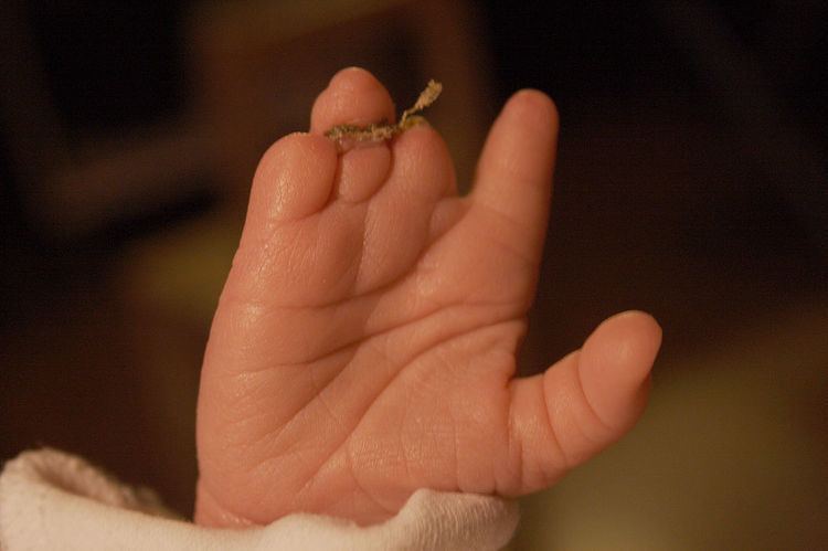 | ||
Constriction ring syndrome (CRS), also known or misnamed as "ADAM Complex", "amniotic band sequence", "amniotic band syndrome", "amniotic disruption complex", "amniochorionic mesoblastic fibrous strings", "congenital amputation", "constriction band syndrome", "congenital constriction bands", "Streeter bands", "tissue bands", is a congenital disorder with unknown etiology. Because of the unknown etiology there are many different, and sometimes wrong names, (see etiology). It is a malformation due to intrauterine bands or rings that give deep grooves in, most commonly, distal extremities like fingers and toes. In rare cases the constriction ring can form around other parts of the fetus and cause amputation or even intrauterine death (see Types and Classification). The anatomy proximal to the site of constriction (or amputation) is developmentally normal. CRS can be associated with other malformations with club foot being most common. (see also Types and Classification). The precise configuration of the bands, lymphedema, and character of the amputations are not predictable and vary with each individual patient. Also more than one extremity is usually affected, and it is rare for only one ring to present as an isolated malformation with no other manifestation of this syndrome.
Contents
Diagnosis
The diagnosis of constriction ring syndrome can be confirmed with an ultrasonography. The clinical manifestations can be extremely variable. It could be a single or multiple manifestation. This can be confirmed at the end of the first trimester or at the beginning of the second trimester. But not every patient will be diagnosed at that moment, most will get this diagnosis at birth.
Differential Diagnosis;
The differential diagnosis includes;
ADAM Complex; CRS is sometimes mislabeled as ADAM complex. ADAM is an abbreviation for Amniotic Deformity, Adhesions Mutilations. CRS is the malformation due to a constriction ring around mostly a limb. ADAM-complex is the association of limb defects (caused by constriction rings) and certain craniofacial clefts
“Adams-Oliver syndrome is often mislabeled as CRS and consists of cutis aplasia of the scalp in which a longitudinal defect can vary in size and can often be associated with full-thickness skullcap loss. The distal digital or toe hypoplasia-aplasia is often confused with CRS. Constriction rings with or without edema are not present. The digital or toe hypoplasia-aplasia usually contains diminutive nails or nail folds”.
Etiology
There are three different theories to the etiology of the Constriction Ring Syndrome.
The first is the intrinsic theory, which was proposed by Streeter in 1930, implicates an anomaly in germ plasm resulting in the defects. This theory is reinforced by the clinical presentation of the constriction rings with other internal visceral and systematic anomalies. Because of these other anomalies the names “Constriction Ring Syndrome”, “Constriction Band Syndrome” and “Streeter Bands” are given to this defect/disease.
The second theory postulates the involvement of an intrauterine disruption during pregnancy followed by a cascade of events involving amniotic rupture. When spontaneous rupture of the amnion occurs early in the second trimester, the separation of amnion from chorion produces many small, thin strands that can become entangled within digits and toes. The names “Amniotic Band Syndrome”, “Amniotic Disruption Complex", "Amniochorionic Mesoblastic Fibrous Strings", are based on this theory.
The third theory postulates the involvement of intrauterine trauma. Intrauterine trauma could be something like amniocentesis, or something like an fetal surgery. An intrauterine trauma could result in hemorrhage leading to acrosyndactyly. One study also showed the presence of bands as confirmed by sonography after fetal surgery.
Because of these different theories, there are many names for this syndrome. For a long time people believed the second theory about the amniotic rupture and strands. In the research cases not every child had a real (amniotic) strand. It could be that there has to be another explanation for the development of these anomalies.
Types & Classification
The constriction ring syndrome is a complex collection of asymmetric congenital anomalies, in which no two cases are exactly alike. This is why a classification is difficult to make.
The most widely used classification system was proposed by Patterson. This classification system is based on the severity of the syndrome and is useful because, the different types require different treatments. Other clinicians have amended this scheme by separating the depth of the ring into mild, moderate, severe and amputation and by further defining the presence or absence of lymphedema or soft tissue loss distal to the ring. Expanding over subdivision in depth of the clefts for every classification is not necessary because the principles of treatment and technique for correction are the same
There are four categories:
- simply constriction rings
- constriction rings associated with deformity of the distal part with or without lymphedema
- constriction rings associated with acrosyndactyly
- uterine amputation
Patterson divided the constriction ring associated with acrosyndactyly into three types:
Patterson I; There are simple constriction rings which are strands most commonly around the distal extremities as fingers and toes. In general, the thumb is not likely to be affected by a constriction ring because the fetus typically holds the thumb in tight adduction flexion, making entanglement with strands less likely. These malformations need to be surgically removed which must be executed in different stages and can done by different techniques (see also treatment).
Patterson II; The CRS involves strands which obstruct the lymphatic vessels and thus causing fluid retention, distal of the affected extremity. This utters itself with swollen parts distal of the constriction.
Patterson III; In this form there is a complex form of syndactyly named acrosyndactyly, the fingers (or toes) were initially separated but due to the constriction they are formed back together. Sometimes multiple fingers can be involved. The distal fusion between digits or toes never initially involves a skeletal coalition. The digits are usually hypoplastic if multiple digits are involved. When the constriction cuts of the blood supply to the fingers, the fingers can form a peak with the most palmar digit being the index finger. Normal neurovascular bundles are not present in the distal parts. Hands with fused fingers need to be released in phases to preserve the distal blood supply.
Paterson IV; One of the most severe consequence of constriction strains is probably intrauterine amputations, this is where the constriction goes as deep as the bone and cuts of the blood supply of the proximal extremity. The result will be that the developing toe or finger will become ischemic and will fall of. Because the end result is a transverse amputation that cuts off the vascular supply to the developing extremity, the actual constriction ring is not seen This can result in different outcomes;
Intrauterine death; In extremely rare cases a strain can form around the umbilical cord and cut off the blood supply to the fetus which will result in intrauterine death.
Malformation associated with constriction ring syndrome; The percentage of associated anomalies varies from 40% to as high as 80% Constriction ring deformities are as common on the lower extremity as on the upper, almost all of these involve the musculoskeletal system, with clubbed feet being the most common in up to 30% of reported cases Large reported series reveal an incidence between 5% and 15% of craniofacial malformations with clefting of the lip or palate.
Epidemiology
The reported incidence of constriction ring syndrome varies from 1/1200 and 1/15000 live births. The prevalence is equally in male and female.
Fetomaternal factors like prematurity, maternal illnes, low birth weight and maternal drug exposure are predisposing factors for the constriction ring syndrome.
No positive relationship between CRS and genetic inheritance has been reported.
Treatment
Surgical correction is recommended when a constriction ring results in a limb contour deformity, with or without lymphedema.
Surgical technique At the beginning of the surgery a tourniquet will be applied to the limb. A tourniquet compresses and control the arterial and venous circulation for about 2 hours. The constriction band must be dissected very carefully to avoid damaging the underlying neurovasculature. When the constriction band is excised, there will be a direct closure. This allows the fatty tissue to naturally reposition itself under the skin.
“With complete circumferential constriction bands, it is recommended that a two-stage correction approach be used. At the first operation, one-half of the circumference is excised and the other one-half can be excised after three to six months. This will avoid any problems to the distal circulation in the limb, which may already be compromised. Lymphedema, when present, will significantly improve within a few weeks of the first surgery.”
For the direct closure of the defect after dissecting a constriction band there are two different techniques:
- Triangular flaps; For this technique the circumference between the two borders must be measured. Depending on the difference the number of triangular flaps can be decided. With a triangular flap you can create more skin.
- Z/W-plasty; “Z-plasty is a plastic surgery technique that is used to improve the functional and cosmetic appearance of scars. It can elongate a contracted scar or rotate the scar tension line. The middle line of the Z-shaped incision (the central element) is made along the line of greatest tension or contraction, and triangular flaps are raised on opposite sides of the two ends and then transposed.”
In rare cases, if diagnosed in utero, fetal surgery may be considered to save a limb which is in danger of amputation or other deformity. This typically would not be attempted if neither vital organs nor the umbilical cord were affected. This operation has been successfully performed on fetuses as young as 22 weeks. The surgery took place at Melbourne's Monash Medical Centre in Australia and is believed to be the earliest surgery of its type, as surgeons usually hold off on operating until the woman is in week 28 of gestation. There are also several facilities in the United States that have performed successful amniotic band release surgery.