
Latin chorion Code TE E5.11.3.1.1.0.3 | MeSH A10.615.284.473 | |
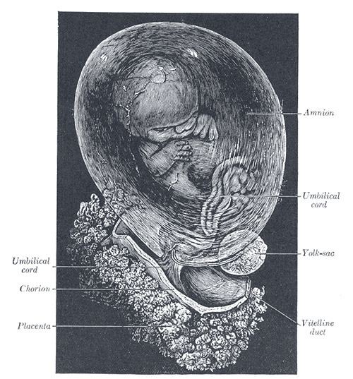 | ||
In humans and most mammals, the chorion is one of the membranes that exist during pregnancy between the developing fetus and mother (the fetal membranes). The chorion and the amnion together form the amniotic sac.
Contents
It is formed by extraembryonic mesoderm and the two layers of trophoblast that surround the embryo and other membranes. The chorionic villi emerge from the chorion, invade the endometrium, and allow transfer of nutrients from maternal blood to fetal blood.
Layers
The chorion consists of two layers: an outer formed by the trophoblast, and an inner formed by the somatic mesoderm; the amnion is in contact with the latter.
The trophoblast is made up of an internal layer of cubical or prismatic cells, the cytotrophoblast or layer of Langhans, and an external layer of richly nucleated protoplasm devoid of cell boundaries, the syncytiotrophoblast.
Growth
The chorion undergoes rapid proliferation and forms numerous processes, the chorionic villi, which invade and destroy the uterine decidua, while simultaneously absorbing nutritive materials from it for the growth of the embryo.
The chorionic villi are at first small and non-vascular, and consist of the trophoblast only, but they increase in size and ramify, whereas the mesoderm, carrying branches of the umbilical vessels, grows into them, and they are vascularized.
Blood is carried to the villi by the paired umbilical arteries, which branch into chorionic arteries and enter the chorionic villi as cotyledon arteries. After circulating through the capillaries of the villi, the blood is returned to the embryo by the umbilical vein. Until about the end of the second month of pregnancy, the villi cover the entire chorion, and are almost uniform in size; but, after this, they develop unequally.
Parts
The part of the chorion that is in contact with the decidua capsularis undergoes atrophy, so that by the fourth month scarcely a trace of the villi is left. This part of the chorion becomes smooth, and is named the chorion laeve (from the Latin word levis, meaning smooth). As it takes no share in the formation of the placenta, this is also named the non-placental part of the chorion. As the chorion grows, the chorion laeve comes in contact with the decidua parietalis and these layers fuse.
On the other hand, the villi at the embryonic pole, which is in contact with the decidua basalis, increase greatly in size and complexity, and hence this part is named the chorion frondosum.
Thus the placenta develops from the chorion frondosum and the decidua basalis.
Monochorionic twins
Monochorionic twins are twins that share the same placenta. It occurs in 0.3% of all pregnancies, and in 75% of monozygotic (identical) twins, when the split takes place on or after the third day after fertilization. The remaining 25% of monozygous twins become dichorionic diamniotic. The condition may affect any type of multiple birth, resulting in monochorionic multiples.