
Specialty urology ICD-9-CM 593.89, 753.23 MedlinePlus 000462 | ICD-10 N28.8 DiseasesDB 33455 eMedicine radio/729 | |
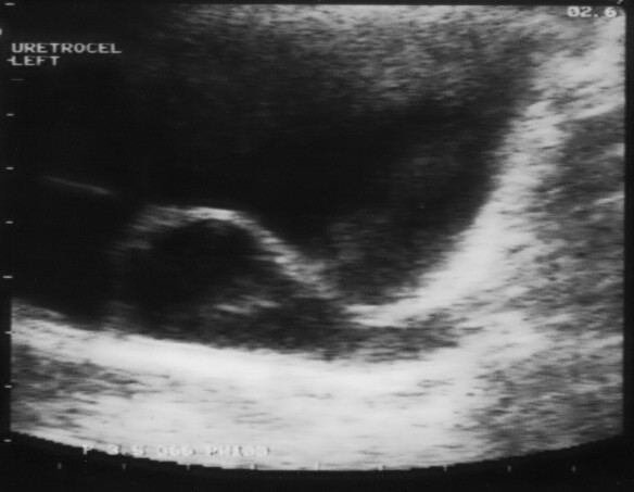 | ||
A ureterocele is a congenital abnormality found in the ureter. In this condition the distal ureter balloons at its opening into the bladder, forming a sac-like pouch. It is most often associated with a duplicated collection system, where two ureters drain their respective kidney instead of one. Simple ureterocele, where the condition involves only a single ureter, represents only twenty percent of cases. Ureterocele affects one in 4,000 individuals, at least four-fifths of whom are female. Patients are frequently Caucasian.
Contents
Since the advent of the ultrasound, most ureteroceles are diagnosed prenatally. The pediatric and adult conditions are often found incidentally, i.e. through diagnostic imaging performed for unrelated reasons.
Classification
Signs and symptoms
The signs and symptoms of ureterocele in the latter two forms can easily be confused with other medical conditions. Symptoms can include:
Complications
Many other complications arise from ureteroceles. Redundant collection systems are usually smaller in diameter than single, and predispose the patient to impassable kidney stones. The effective "bladder within a bladder" compounds this problem by increasing the collision of uric acid particles, the process by which uric acid stones are formed. Ureterocele is also associated with poor kidney function. It can cause frequent blockage of the ureter leading to serious kidney damage. In other cases, a small, upper portion of the kidney is congenitally non-functional. Though often benign, this problem can necessitate the removal of non-functioning parts.
Causes
Definitive causes of ureterocele have not been found. While the abnormal growth occurs within the uterus, it has not been substantiated that genetics are to blame.