1835French dermatologist
Pierre François Olive Rayer published an atlas of skin diseases. It contains 22 large coloured plates with 400 figures presented in a systematic order. On page 20, fig. 1 is a drawing that is regarded as the earliest description of tuberous
sclerosis. Entitled
"végétations vasculaires", Rayer noted these were "small vascular, of papulous appearance, widespread growths distributed on the nose and around the mouth". No mention was made of any medical condition associated with the skin disorder.
1850English dermatologists
Thomas Addison and William Gull described, in
Guy's Hospital Reports, the case of a four-year-old girl with a "peculiar eruption extending across the nose and slightly affecting both cheeks", which they called "vitiligoidea tuberosa".
1862German physician
Friedrich Daniel von Recklinghausen, who was working as an assistant to
Rudolf Virchow in the Institute for Pathological Anatomy in Berlin, presented a case to the city's Obstetrical Society. The heart of an infant who "died after taking a few breaths" had several tumours. He called these tumours "myomata", one of which was the "size of a pigeon's egg". He also noted the brain had "a great number of scleroses". These were almost certainly the cardiac
rhabdomyomas and cortical tubers of tuberous sclerosis. He failed to recognise a distinct disease, regarding it as a pathological-anatomical curiosity. Von Recklinghausen's name would instead become associated with
neurofibromatosis after a classic paper in 1881.
1864German pathologist
Rudolf Virchow published a three-volume work on tumours that described a child with cerebral tuberous sclerosis and rhabdomyoma of the heart. His description contained the first hint that this may be an inherited disease: the child's sister had died of a cerebral tumour.
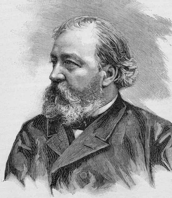
1880French neurologist
Désiré-Magloire Bourneville had a chance encounter with the disease that would bear his name. He was working as an unofficial assistant to Jean Martin Charcot at La Salpêtrière. While substituting for his teacher,
Louis J.F. Delasiauve, he attended to Marie, a 15-year-old girl with
psychomotor retardation,
epilepsy and a "confluent vascular-papulous eruption of the nose, the cheeks and forehead". She had a history of seizures since infancy and was taken to the children's hospital aged three and declared a hopeless case. She had learning difficulties and could neither walk nor talk. While under Bourneville's care, Marie had an ever-increasing number of seizures, which came in clusters. She was treated with
quinquina,
bromide of
camphor,
amyl nitrite, and the application of
leeches behind the ears. On 7 May 1879 Marie died in her hospital bed. The post-mortem examination disclosed hard, dense tubers in the cerebral convolutions, which Bourneville named
Sclérose tubéreuse des circonvolutions cérébrales. He concluded they were the source (focus) of her seizures. In addition, whitish hard masses, one "the size of a walnut", were found in both kidneys.
1881German physician Hartdegen described the case of a two-day-old baby who died in
status epilepticus. Post-mortem examination revealed small tumours in the
lateral ventricles of the brain and areas of cortical sclerosis, which he called "glioma gangliocellulare cerebri congenitum".
1881Bourneville and Édouard Brissaud examined a four-year-old boy at
La Bicêtre. As before, this patient had cortical tubers, epilepsy and learning difficulties. In addition he had a
heart murmur and, on post-mortem examination, had tiny hard tumours in the ventricle walls in the brain (subependymal nodules) and small tumours in the kidneys (
angiomyolipomas).
1885French physicians
Félix Balzer and
Pierre Eugène Ménétrier reported a case of
"adénomes sébacés de la face et du cuir" (adenoma of the sebaceous glands of the face and scalp). The term has since proved to be incorrect as they are neither adenoma nor derived from sebaceous glands. The papular rash is now known as facial angiofibroma.
1885French dermatologists
François Henri Hallopeau and Émile Leredde published a case of adenoma sebaceum that was of a hard and fibrous nature. They first described the
shagreen plaques and later would note an association between the facial rash and epilepsy.
1890Scottish dermatologist
John James Pringle, working in London, described a 25-year-old woman with subnormal intelligence, rough lesions on the arms and legs, and a papular facial rash. Pringle brought attention to five previous reports, two of which were unpublished. Pringle's adenoma sebaceum would become a common
eponym for the facial rash.
1901Italian physician GB Pellizzi studied the pathology of the cerebral lesions. He noted their dysplastic nature, the cortical
heterotopia and defective myelination. Pellizzi classified the tubers into type 1 (smooth surface) and type 2 (with central depressions).
1903German physician Richard Kothe described periungual fibromas, which were later rediscovered by the Dutch physician Johannes Koenen in 1932 (known as Koenen's tumours).
1906Australian neurologist
Alfred Walter Campbell, working in England, considered the lesions in the brain, skin, heart and kidney to be caused by one disease. He also first described the pathology in the eye. His review of 20 reported cases led him to suggest a diagnostic triad of symptoms that is more commonly attributed to Vogt.
1908German paediatric neurologist Heinrich Vogt established the diagnostic criteria for TSC, firmly associating the facial rash with the neurological consequences of the cortical tubers. Vogt's triad of epilepsy, idiocy, and adenoma sebaceum held for 60 years until research by
Manuel Gómez discovered that fewer than a third of patients with TSC had all three symptoms.
1910J. Kirpicznick was first to recognise that TSC was a genetic condition. He described cases of identical and fraternal twins and also one family with three successive generations affected.
1911Edward Sherlock, barrister-at-law and lecturer in biology, reported nine cases in his book on the "feeble-minded". He coined the term
epiloia, a portmanteau of epilepsy and anoia (mindless). The word is no longer widely used as a synonym for TSC. The geneticist Robert James Gorlin suggested in 1981 that it could be a useful acronym for
epilepsy,
low
intelligence, and
adenoma sebaceum.
1913H. Berg is credited with first stating that TSC was a hereditary disorder, noting its transmission through two or three generations.
1914P. Schuster described a patient with adenoma sebaceum and epilepsy but of normal intelligence. This reduced phenotypic expression is called a
forme fruste.
1918French physician René Lutembacher published the first report of cystic lung disease in a patient with TSC. The 36-year-old woman died from bilateral pneumothoraces. Lutembacher believed the cysts and nodules to be metastases from a renal
fibrosarcoma. This complication, which only affects women, is now known as
lymphangioleiomyomatosis (LAM).
1920Dutch ophthalmologist
Jan van der Hoeve described the
retinal hamartomas (phakoma). He grouped both TSC and
neurofibromatosis together as "phakomatoses" (later called neurocutaneous syndromes).
1924H. Marcus noted that characteristic features of TSC such as intracranial calcifications were visible on
x-ray.
1932MacDonald Critchley and Charles J.C. Earl studied 29 patients with TSC who were in mental institutions. They described behaviour—unusual hand movements, bizarre attitudes and repetitive movements (stereotypies)—that today would be recognised as autistic. However it would be 11 years before
Leo Kanner suggested the term "
autism". They also noticed the associated white spots on the skin (hypomelanic macules).
1934N.J. Berkwitz and L.G. Rigler showed it was possible to diagnose tuberous sclerosis using
pneumoencephalography to highlight non-calcified subependymal nodules. These resembled "the wax drippings of a burning candle" on the lateral ventricles.
1942Sylvan E. Moolten proposed "the tuberous sclerosis complex", which is now the preferred name. This recognises the multi-organ nature of the disease. Moolten introduced three words to describe its pathology: "the basic lesion is
hamartial, becoming in turn tumor-like (
hamartoma) or truly neoplastic (
hamartoblastoma)."
1954Norwegian pathologist Reidar Eker bred a line of Wistar rats predisposed to renal
adenomas. The Eker rat became an important model of dominantly inherited cancer.
1966Phanor Perot and Bryce Weir pioneered surgical intervention for epilepsy in TSC. Of the seven patients who underwent cortical tuber resection, two became seizure-free. Prior to this, only four patients had ever been surgically treated for epilepsy in TSC.
1967J.C. Lagos and
Manuel Rodríguez Gómez reviewed 71 TSC cases and found that 38% of patients have normal intelligence.
1971American geneticist
Alfred Knudson developed his "two hit" hypothesis to explain the formation of
retinoblastoma in both children and adults. The children had a congenital
germline mutation which was combined with an early lifetime
somatic mutation to cause a tumour. This model applies to many conditions involving tumour suppressor genes such as TSC. In the 1980s, Knudson's studies on the Eker rat strengthened this hypothesis.
1975Giuseppe Pampiglione and E. Pugh, in a letter to
The Lancet, noted that up to 69% of patients presented with infantile spasms.
1975Riemann first used ultrasound to examine TSC-affected kidneys in the case of a 35-year-old woman with chronic renal failure.
1976Cranial computed tomography (CT, invented 1972) proved to be an excellent tool for diagnosing cerebral neoplasms in children, including those found in tuberous sclerosis.
1979Manuel Gómez published a
monograph:
"Tuberous Sclerosis" that remained the standard textbook for three editions over two decades. The book described the full clinical spectrum of TSC for the first time and established a new set of diagnostic criteria to replace the Vogt triad.
1982Kenneth Arndt successfully treated facial angiofibroma with an argon laser.
1983Positron emission tomography (PET, invented 1981) was compared to
electroencephalography (EEG) and CT. It was found to be capable of locating epileptogenic cortical tubers that would otherwise have been missed.
1984The cluster of infantile spasms in TSC was discovered to be preceded by a focal EEG discharge.
1985Magnetic resonance imaging (MRI, invented 1980) was first used in TSC to identify affected regions in the brain of a girl with tuberous sclerosis.
1987MR was judged superior to CT imaging for both sensitivity and specificity. In a study of fifteen patients, it identified subependymal nodules projecting into the lateral ventricles in twelve patients, distortion of the normal cortical architecture in ten patients (corresponding to cortical tubers), dilated ventricles in five patients, and distinguished a known
astrocytoma from benign subependymal nodules in one patient.
1987MR imaging was found to be capable of predicting the clinical severity of the disease (epilepsy and developmental delay). A study of 25 patients found a correlation with the number of cortical tubers identified. In contrast, CT was not a useful predictor, but was superior at identifying calcified lesions.
1987Linkage analysis on 19 families with TSC located a probable gene on chromosome 9.
1988Cortical tubers found on MR imaging corresponded exactly to the location of persistent EEG foci, in a study of six children with TSC. In particular, frontal cortical tubers were associated with more intractable seizures.
1990Vigabatrin was found to be a highly effective antiepileptic treatment for infantile spasms, particularly in children with TSC. Following the discovery in 1997 of severe persistent
visual field constriction as a possible side-effect, vigabatrin monotherapy is now largely restricted to this patient group.
1992Linkage analysis located a second gene to chromosome 16p13.3, close to the
polycystic kidney disease type 1 (PKD1) gene.
1993The European Chromosome 16 Tuberous Sclerosis Consortium announced the cloning of
TSC2; its product is called tuberin.
1994The Eker rat was discovered to be an animal model for tuberous sclerosis; it has a mutation in the rat-equivalent of the TSC2 gene.
1995MRI with fluid attenuated inversion recovery (FLAIR) sequences was reported to be significantly better than standard T
2-weighted images at highlighting small tubers, especially subcortical ones.
1997The TSC1 Consortium announced the cloning of TSC1; its product is called hamartin.
1997The PKD1 gene, which leads to autosomal dominant polycystic kidney disease (ADPKD), and the TSC2 gene were discovered to be adjacent on chromosome 16p13.3. A team based at the Institute of Medical Genetics in Wales studied 27 unrelated patients with TSC and renal cystic disease. They concluded that serious renal disease in those with TSC is usually due to contiguous gene deletions of TSC2 and PKD1. They also noted that the disease was different (earlier and more severe) than ADPKD and that patients with TSC1 did not suffer significant cystic disease.
1997Patrick Bolton and Paul Griffiths examined 18 patients with TSC, half of whom had some form of autism. They found a strong association between tubers in the temporal lobes and the patients with autism.
1998The Tuberous Sclerosis Consensus Conference issued revised diagnosic criteria, which is the current standard.
1998An Italian team used
magnetoencephalography (MEG) to study three patients with TSC and partial epilepsy. Combined with MRI, they were able to study the association between tuberous areas of the brain, neuronal malfunctioning and epileptogenic areas. Later studies would confirm that MEG is superior to EEG in identifying the eliptogenic tuber, which may be a candidate for surgical resection.
2001A multi-centre
cohort of 224 patients were examined for mutations and disease severity. Those with TSC1 were less severely affected than those with TSC2. They had fewer seizures and less mental impairment. Some symptoms of TSC were rare or absent in those with TSC1. A conclusion is that "both germline and somatic mutations appear to be less common in TSC1 than in TSC2".
2002Several research groups investigated how the TSC1 and TSC2 gene products (tuberin and hamartin) work together to inhibit mammalian target of rapamycin (mTOR)-mediated downstream signalling. This important pathway regulates cell proliferation and tumour suppression.
2002Treatment with rapamycin (sirolimus) was found to shrink tumours in the Eker rat (TSC2) and mouse (TSC1) models of tuberous sclerosis.
2006Small trials showed promising results in the use of rapamycin to shrink angiomyolipoma and astrocytomas. Several larger multicentre clinical trials began: lymphangioleiomyomatosis (LAM) and kidney angiomyolipoma (AML) were treated with rapamycin; giant cell astrocytomas were treated with the rapamycin derivative
everolimus.