
Latin Lamina terminalis NeuroNames hier-190 FMA 61975 | MeSH A08.186.211.577.482 TA A14.1.08.419 | |
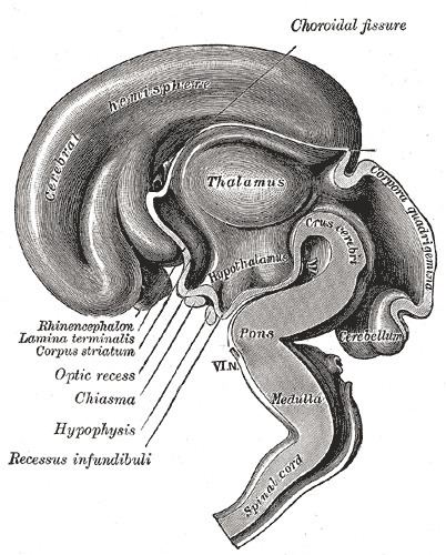 | ||
The median portion of the wall of the fore-brain vesicle consists of a thin lamina, the lamina terminalis, which stretches from the Interventricular foramen (Foramen of Monro) to the recess at the base of the optic stalk and contains the organum vasculosum of the lamina terminalis, which regulates the osmolarity of the blood. The lamina terminalis is the anterior version of the tuber cinereum, together they form the infundibulum.
The lamina terminalis can be opened via endoscopic neurosurgery in an attempt to create a path that cerebrospinal fluid can flow through when a person suffers from hydrocephalus and when it is not possible to perform an Endoscopic third ventriculostomy, but the effectiveness of this technique is not certain.
This is the rostral end (tip) of the neural tube (embryological central nervous system) in the early weeks of development. Failure of the lamina terminalis to close properly at this stage of development will result in anencephaly or meroencephaly.