
Latin Canalis tarsi | Dorlands/Elsevier 12832517 | |
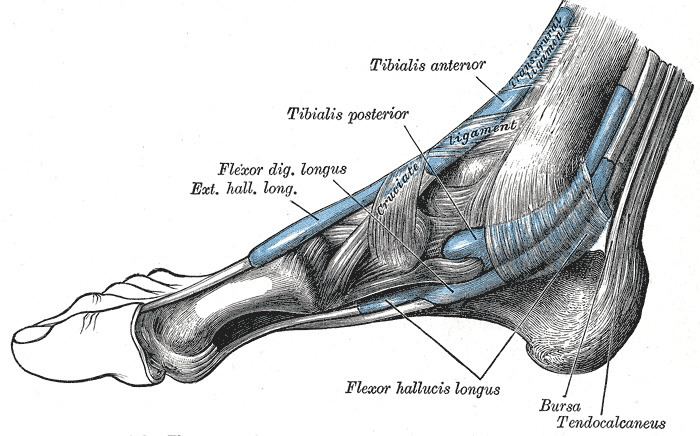 | ||
The tarsal tunnel is found along the inner leg posterior to the medial malleolus.
Contents
Structure
The tarsal tunnel is made up of bone on the inside and the flexor retinaculum on the outside.
Contents
The tibial nerve, posterior tibial artery, veins, and tendons travel in a bundle along this pathway, through the tarsal tunnel, as do:
In the tunnel, the nerve splits into three different paths. One nerve (calcaneal) continues to the heel, the other two (medial plantar nerve and lateral plantar nerve) continue on to the bottom of the foot.
Tarsal tunnel syndrome
Tarsal tunnel syndrome is the most commonly reported nerve entrapment of the ankle and is analogous to the carpal tunnel of the wrist. People with tarsal tunnel syndrome have pain in the plantar aspect of the foot mostly at night. Weight bearing increases pain and weakness is found on intrinsic foot muscles with positive Tinel sign at the tunnel. There is no tenderness present on the plantar foot, though this is typically the primary site of complaint.