
 | ||
Antagonist Fibularis brevis and longus, antagonist to the inversion. | ||
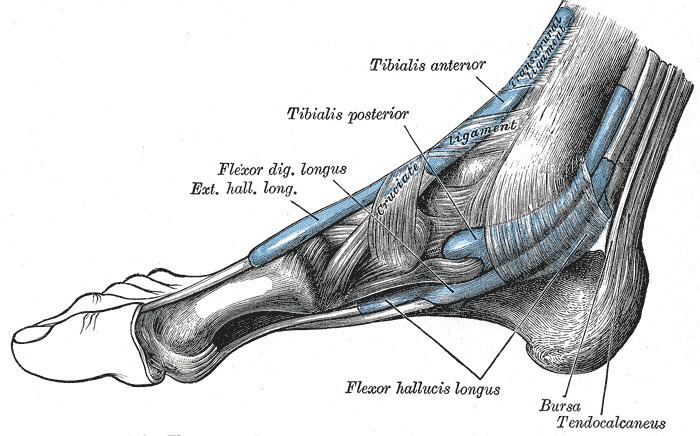
The tibialis posterior is the most central of all the leg muscles, and is located in the deep posterior compartment of the leg.
Contents
It is the key stabilizing muscle of the lower leg.
Blood is supplied to the muscle by the posterior tibial artery, and innervation is via the tibial nerve.
Structure
The tibialis posterior muscle originates on the inner posterior borders of the tibia and fibula. It is also attached to the interosseous membrane, which attaches to the tibia and fibula.
The tendon of the tibialis posterior muscle (sometimes called the posterior tibial tendon) descends posterior to the medial malleolus and terminates by dividing into plantar, main, and recurrent components. The plantar portion inserts into the bases of the second, third and fourth metatarsals, the intermediate and lateral cuneiforms and the cuboid. The main portion inserts into the tuberosity of the navicular and the plantar surface of the medial cuneiform. The recurrent portion inserts into the sustentaculum tali of the calcaneus.
Function
As well as being a key muscle and tendon for stabilization, the tibialis posterior also contracts to produce inversion and assists in the plantar flexion of the foot at the ankle. The tibialis posterior has a major role in supporting the medial arch of the foot. Dysfunction of the tibialis posterior, including rupture of the tibialis posterior tendon, can lead to flat feet in adults, as well as a valgus deformity due to unopposed eversion when inversion is lost.