
 | ||
Nerve tibial nerve, S1 & S2 more than L5 nerve roots Antagonist | ||
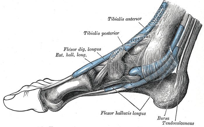
The Flexor hallucis longus muscle (FHL) is one of the three deep muscles of the posterior compartment of the leg that distally attaches to the plantar surface of the distal phalanx of the hallux (great or big toe). The other deep muscles are the flexor digitorum longus and tibialis posterior; the tibialis posterior is the most powerful of these deep muscles. All three muscles are innervated by the tibial nerve which comprises half of the sciatic nerve.
Contents
Structure
The Flexor hallucis longus is situated on the fibular side of the leg. It arises from the inferior two-thirds of the posterior surface of the body of the fibula, with the exception of 2.5 cm. at its lowest part; from the lower part of the interosseous membrane; from an intermuscular septum between it and the Peronæi, laterally, and from the fascia covering the Tibialis posterior, medially.
The fibers pass obliquely downward and backward, where it passes through the tarsal tunnel on the medial side of the foot and end in a tendon which occupies nearly the whole length of the posterior surface of the muscle.
This tendon lies in a groove which crosses the posterior surface of the lower end of the tibia, between the medial and lateral tubercoles of the posterior surface of the talus, and the under surface of the sustentaculum tali of the calcaneus; in the sole of the foot it runs forward between the two heads of the Flexor hallucis brevis, and is inserted into the base of the last phalanx of the great toe. The grooves on the talus and calcaneus, which contain the tendon of the muscle, are converted by tendinous fibers into distinct canals, lined by a mucous sheath.
As the tendon passes forward in the sole of the foot, it is situated above, and crosses from the lateral to the medial side of the tendon of the Flexor digitorum longus, to which it is connected by a fibrous slip.
Variation
Usually a slip runs to the Flexor digitorum and frequently an additional slip runs from the Flexor digitorum to the Flexor hallucis. Peroneocalcaneus internus, rare, arises below or outside the Flexor hallucis from the back of the fibula, passes over the sustentaculum tali with the Flexor hallucis and inserts into the calcaneum.
Function
Similar to the flexor digitorum longus and tibialis posterior muscles, the flexor hallucis longus muscle functions to plantar flex and invert the foot. However, it is unique in that it also functions to flex the great toe and helps supinate the ankle.
Injury and Treatment
Common injuries associated with the FHL tendon are tenosynovitis, tendinopathies, and muscle strains. After passing through the tarsal tunnel, the flexor hallucis longus tendon must curve around a bony landmark called the sustenaculum tali. Friction at this site is likely to cause pain on the posteromedial aspect of the ankle. While commonly referred to as "dancer's tendinitis," FHL tendinitis occurs commonly in both ballet dancers and runners. Due to their excessive use of toe flexion, inflammation and irritation is common at the site of the sustenaculum tali. Hallux saltans is a condition that develops as a result of overusing the FHL muscle. With this condition, a nodule develops along the FHL tendon which may produce a popping effect during contraction because it drags along surrounding tissues.
Some FHL injuries can be treated through rest, physical therapy, splints, and anti-inflammatory medication. However, more serious or chronic injuries may require surgery.