
 | ||
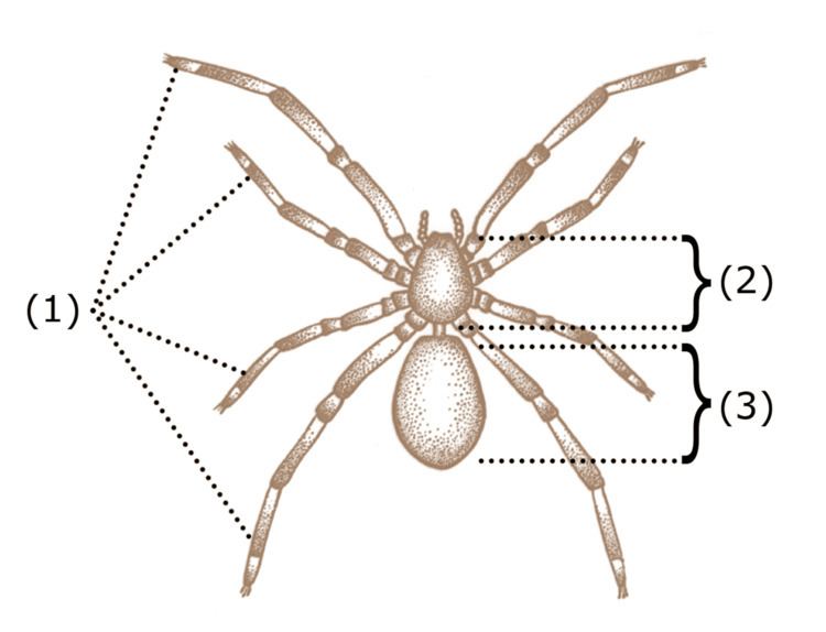
The anatomy of spiders includes many characteristics shared with other arachnids. These characteristics include bodies divided into two tagmata (sections or segments), eight jointed legs, no wings or antennae, the presence of chelicerae and pedipalps, simple eyes, and an exoskeleton, which is periodically shed.
Contents
Spiders also have several adaptations that distinguish them from other arachnids. All spiders are capable of producing silk of various types, which many species use to build webs to ensnare prey. Most spiders possess venom, which is injected into prey (or defensively, when the spider feels threatened) through the fangs of the chelicerae. Male spiders have specialized pedipalps that are used to transfer sperm to the female during mating. Many species of spiders exhibit a great deal of sexual dimorphism.
Circulation
Spiders, like most arthropods, have an open circulatory system, i.e., they do not have true blood, or veins to convey it. Rather, their bodies are filled with haemolymph, which is pumped through arteries by a heart into spaces called sinuses surrounding their internal organs. The haemolymph contains hemocyanin, a respiratory protein similar in function to hemoglobin. Hemocyanin contains two copper atoms, tinting the haemolymph with a faint blue color.
The heart is located in the abdomen a short distance within the middle line of the dorsal body-wall, and above the intestine. Unlike in insects, the heart is not divided into chambers, but consists of a simple tube. The aorta, which supplies haemolymph to the cephalothorax, extends from the anterior end of the heart. Smaller arteries extend from sides and posterior end of the heart. A thin-walled sac, known as the pericardium, completely surrounds the heart.
Breathing
Spiders have developed several different respiratory anatomies, based either on book lungs or on tracheae. Mesothele and mygalomorph spiders have two pairs of book lungs filled with haemolymph, where openings on the ventral surface of the abdomen allow air to enter and oxygen to diffuse in and carbon dioxide to diffuse out. This is also the case for some basal araneomorph spiders like the family Hypochilidae, but the remaining members of this group have just the anterior pair of book lungs intact while the posterior pair of breathing organs are partly or fully modified into tracheae, through which oxygen is diffused into the haemolymph or directly to the tissue and organs. This system has most likely evolved in small ancestors to help resist desiccation. The trachea were originally connected to the surroundings through a pair of spiracles, but in the majority of spiders this pair of spiracles has fused into a single one in the middle, and migrated posterior close to the spinnerets.
Among smaller araneomorph spiders there are species in which the anterior pair of book lungs have also evolved into tracheae, or are simply reduced or missing. In a very few species the book lungs have developed deep channels, apparently signs of evolution into tracheae. Some very small spiders in moist and sheltered habitats do not have any breathing organs at all, as gas exchange occurs directly through their body surface. In the tracheal system oxygen interchange is much more efficient, enabling cursorial hunting (hunting involving extended pursuit) and other advanced characteristics, such as having a smaller heart and the ability to live in drier habitats.
Digestion
Digestion is carried out internally and externally. Spiders do not have powerful chelicerae, but secrete digestive fluids into their prey from a series of ducts perforating their chelicerae. The coxal glands are excretory organs that lie in the prosoma, and open to the outside at the coxae of the walking legs. In primitive spiders, such as the Mesothelae and the Mygalomorphae, two pairs of coxal glands open onto the posterior side of the first and third coxae. They release a fluid only during feeding and play an important role in ion and water balance. Digestive fluids dissolve the prey's internal tissues. Then the spider feeds by sucking the partially digested fluids out. Other spiders with more powerfully built chelicerae masticate the entire body of their prey and leave behind only a relatively small amount of indigestible materials. Spiders consume only liquid foods. Many spiders will store prey temporarily. Web weaving spiders that have made a shroud of silk to quiet their envenomed prey's death struggles will generally leave them in these shrouds and then consume them at their leisure.
Reproductive system
Almost all spiders reproduce sexually. They are unusual in that they do not transfer sperm directly, for example via a penis. Instead the males transfer it to specialized structures (palpal bulbs) on the pedipalps and then meander about to search for a mate. These palps are then introduced into the female's epigyne. This was first described in 1678 by Martin Lister. In 1843 it was revealed that males build a nuptial web into which they deposit a drop of semen, which is then taken up by the copulatory apparatus (the palpal bulb) in the pedipalp. The structure of the copulatory apparatus varies significantly between males of different species. While the widened palpal tarsus of Filistata hibernalis (Filistatidae) only forms a simple bulb containing the coiled blind duct, members of the genus Argiope have a highly complex structure.