
Subclass Digenea Family Schistosomatoidea Rank Species | Order Prosostomata Higher classification Blood-flukes | |
 | ||
Similar Schistosoma mansoni, Schistosoma japonicum, Bulinus, Schistosoma mekongi, Biomphalaria | ||
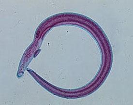
Schistosoma haematobium
Schistosoma haematobium is an important digenetic trematode, and is found in Africa and the Middle East. It is a major agent of schistosomiasis; more specifically, it is associated with urinary schistosomiasis.
Contents
- Schistosoma haematobium
- Life cycle
- Geographical distribution
- Diagnosis
- Prevention
- Immunopathology
- Pathology
- Treatment
- References
Adults are found in the venous plexuses around the urinary bladder and the released eggs travels to the wall of the urine bladder causing haematuria and fibrosis of the bladder. The bladder becomes calcified, and there is increased pressure on ureters and kidneys otherwise known as hydronephrosis. Inflammation of the genitals due to S. haematobium may contribute to the propagation of HIV. Studies have shown the relationship between S. haematobium infection and the development of squamous cell carcinoma of the bladder.
Schistosoma haematobium
Life cycle
The free swimming infective larval cercariae burrow into human skin when it comes into contact with contaminated water. The cercariae enter the blood stream of the host where they travel to the liver to mature into adult flukes. In order to avoid detection by the immune system inside the host, the adults have the ability to coat themselves with host antigen After a period of about three weeks the young flukes migrate to the urinary bladder veins to copulate. The female fluke lays as many as 30 eggs per day which migrate to the lumen of the urinary bladder and ureters. The eggs are eliminated from the host into the water supply with micturition. In fresh water, the eggs hatch forming free swimming miracidia which penetrate into the intermediate snail host (Bulinus spp., e.g. B. globosus, B. forskalii, B. nyassanus and B. truncatus). Inside the snail, the miracidium sheds it epithelium and develops into a mother sporocyst. After two weeks the mother begins forming daughter sporocysts. Four weeks after the initial penetration of the miracidium into the snail furcocercous cercariae begin to be released. The cercariae cycle from the top of the water to the bottom for three days in the search of a human host. Within half an hour the cercariae enter the host epithelium.
Geographical distribution
S. hematobium infects snails in Africa and the Middle East.
Diagnosis
Traditionally, diagnoses has been made by examination of the urine for eggs. In chronic infections, or if eggs are difficult to find, an intradermal injection of schistosome antigen to form a wheal is effective in determining infection. Alternatively diagnosis can be made by complement fixation tests, As of 2012 commercial serologic tests included ELISA and an Indirect immunofluorescence test, hampered by a low sensitivity ranging from 21% to 71%.
Prevention
The main cause of schistomiasis is the dumping of human waste into water supplies. Hygienic disposal of waste would be sufficient to eliminate the disease.
Immunopathology
The immune system responds to eggs in liver causing hypersensitivity; an immune response is necessary to prevent damage to hepatocytes. The hosts' antibodies which bind to the tegument of the Schistosome don't bind for long since the tegument is shed every few hours. The schistosome can also take on host proteins. Schistomiasis can be divided into three phases: (1) the migratory phase lasting from penetration to maturity,(2) the acute phase which occurs when the schistosomes begin producing eggs, and (3) the chronic phase which occurs mainly in endemic areas.
Pathology
The ova are initially deposited in the muscularis propria which leads to ulceration of the overlaying tissue. Infections are characterized by pronounced acute inflammation, squamous metaplasia, blood and reactive epithelial changes. Granulomas and multinucleated giant cells may be seen.
In late stage the infection may lead to extra-urinary complication named Bilharzial cor pulmonale.
Treatment
The drug of choice is praziquantel, a quinolone derivative.