
Specialty Infectious disease ICD-9-CM 120 | ICD-10 B65 DiseasesDB 11875 1188211856 | |
 | ||
Pronunciation /ˌʃistəsəˈmaɪəsᵻs, -toʊ-, -soʊ-/ | ||
Schistosomiasis, also known as snail fever and bilharzia, is a disease caused by parasitic flatworms called schistosomes. The urinary tract or the intestines may be infected. Signs and symptoms may include abdominal pain, diarrhea, bloody stool, or blood in the urine. Those who have been infected a long time may experience liver damage, kidney failure, infertility, or bladder cancer. In children, it may cause poor growth and learning difficulty.
Contents
- Signs and symptoms
- Intestinal schistosomiasis
- Dermatitis
- Katayama fever
- Chronic disease
- Genitourinary disease
- Gastrointestinal disease
- Central nervous system disease
- Transmission
- Identification of eggs in stools
- Antibody detection
- Prevention
- Snails dams and prawns
- Treatment
- Epidemiology
- Infection estimates
- Deaths
- History
- Society and culture
- Vaccine
- References
The disease is spread by contact with fresh water contaminated with the parasites. These parasites are released from infected freshwater snails. The disease is especially common among children in developing countries as they are more likely to play in contaminated water. Other high risk groups include farmers, fishermen, and people using unclean water during daily living. It belongs to the group of helminth infections. Diagnosis is by finding eggs of the parasite in a person's urine or stool. It can also be confirmed by finding antibodies against the disease in the blood.
Methods to prevent the disease include improving access to clean water and reducing the number of snails. In areas where the disease is common, the medication praziquantel may be given once a year to the entire group. This is done to decrease the number of people infected and, consequently, the spread of the disease. Praziquantel is also the treatment recommended by the World Health Organization (WHO) for those who are known to be infected.
Schistosomiasis affected almost 210 million people worldwide as of 2012. An estimated 12,000 to 200,000 people die from it each year. The disease is most commonly found in Africa, as well as Asia and South America. Around 700 million people, in more than 70 countries, live in areas where the disease is common. In tropical countries, schistosomiasis is second only to malaria among parasitic diseases with the greatest economic impact. Schistosomiasis is listed as a neglected tropical disease.
Signs and symptoms
Many individuals do not experience symptoms. If symptoms do appear, they usually take from four to six weeks from the time of infection. The first symptom of the disease may be a general feeling of illness. Within twelve hours of infection, an individual may complain of a tingling sensation or light rash, commonly referred to as "swimmer's itch", due to irritation at the point of entrance. The rash that may develop can mimic scabies and other types of rashes. Other symptoms can occur two to ten weeks later and can include fever, aching, a cough, diarrhea, chills or gland enlargement. These symptoms can also be related to avian schistosomiasis, which does not cause any further symptoms in humans.
The manifestations of schistosomal infection vary over time as the cercariae, and later adult worms and their eggs migrate through the body. If eggs migrate to the brain or spinal cord, seizures, paralysis, or spinal cord inflammation are possible.
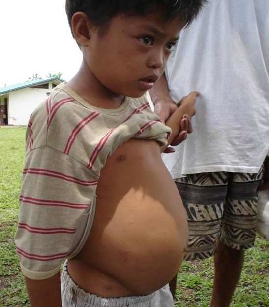
Intestinal schistosomiasis
In intestinal schistosomiasis, eggs become lodged in the intestinal wall and cause an immune system reaction called a granulomatous reaction. This immune response can lead to obstruction of the colon and blood loss. The infected individual may have what appears to be a potbelly. Eggs can also become lodged in the liver, leading to high blood pressure through the liver, enlarged spleen, the buildup of fluid in the abdomen, and potentially life-threatening dilations or swollen areas in the esophagus or gastrointestinal tract that can tear and bleed profusely (esophageal varices). In rare instances, the central nervous system is affected. Individuals with chronic active schistosomiasis may not complain of typical symptoms.
Dermatitis
The first potential reaction is an itchy, papular rash that results from cercariae penetrating the skin, often in a person's first infection. The round bumps are usually one to three centimeters big. Because people living in affected areas have often been repeatedly exposed, acute reactions are more common in tourists and migrants. The rash can occur between the first few hours and a week after exposure and lasts for several days. A similar, more severe reaction called "swimmer's itch" reaction can also be caused by cercariae from animal trematodes that often infect birds.
Katayama fever
Another primary condition, called Katayama fever, may also develop from infection with these worms, and it can be very difficult to recognize. Symptoms include fever, lethargy, the eruption of pale temporary bumps associated with severe itching (urticarial) rash, liver and spleen enlargement, and bronchospasm.
Acute schistosomiasis (Katayama fever) may occur weeks or months after the initial infection as a systemic reaction against migrating schistosomulae as they pass through the bloodstream through the lungs to the liver. Similarly to swimmer's itch, Katayama fever is more commonly seen in people with their first infection such as migrants and tourists. However it is seen in native residents of China infected with S. japonicum. Symptoms include:
The symptoms usually get better on their own but a small proportion of people have persistent weight loss, diarrhea, diffuse abdominal pain and rash.
Chronic disease
In long established disease adult worms lay eggs that can cause inflammatory reactions. The eggs secrete proteolytic enzymes that help them migrate to the bladder and intestines to be shed. However the enzymes also cause an eosinophilic inflammatory reaction when eggs get trapped in tissues or embolize to the liver, spleen, lungs, or brain. The long term manifestations are dependent on the species of schistosome as the adult worms of different species migrate to different areas. Many infections are mildly symptomatic, with anemia and malnutrition being common in endemic areas.
Genitourinary disease
The worms of S. haematobium migrate to the veins around the bladder and ureters. This can lead to blood in the urine 10 to 12 weeks after infection. Over time, fibrosis can lead to obstruction of the urinary tract, hydronephrosis and kidney failure. Bladder cancer diagnosis and mortality are generally elevated in affected areas, and efforts to control schistosomiasis in Egypt have led to decreases in the bladder cancer rate. The risk of bladder cancer appears to be especially high in male smokers, perhaps due to chronic irritation of the bladder lining allowing it to be exposed to carcinogens from smoking.
In women, genitourinary disease can also include genital lesions that may lead to increased rates of HIV transmission.
Gastrointestinal disease
The worms of S. mansoni and S. japonicum migrate to the veins of the gastrointestinal tract and liver. Eggs in the gut wall can lead to pain, blood in the stool, and diarrhea (especially in children). Severe disease can lead to narrowing of the colon or rectum. Eggs also migrate to the liver leading to fibrosis in 4 to 8 percent of people with chronic infection, mainly those with long term heavy infection.
Central nervous system disease
Central nervous system lesions occur occasionally. Cerebral granulomatous disease may be caused by S. japonicum eggs in the brain. Communities in China affected by S. japonicum have been found to have rates of seizures eight times higher than baseline. Similarly, granulomatous lesions from S. mansoni and S. haematobium eggs in the spinal cord can lead to transverse myelitis with flaccid paraplegia. Eggs are thought to travel to the central nervous system via embolization.
Transmission
Infected individuals release Schistosoma eggs into water via their fecal material or urine. After larvae hatch from these eggs, the larvae infect a very specific type of freshwater snail. These larvae undergo the next phase of their lifecycle in these snails, spending their time reproducing and developing. Once this step has been completed, the parasite leaves the snail and enters the water column. The parasite can live in the water for only 48 hours without a human host. Once a host has been found, the worm enters its blood vessels. For several weeks, the worm will remain in the vessels, continuing its development into its adult phase. When maturity is reached, mating occurs and eggs are produced. Eggs enter the bladder/intestine and are excreted through urine and feces and the process repeats. If the eggs do not get excreted, they can become engrained in the body tissues and cause a variety of problems such as immune reactions and organ damage.
Humans encounter larvae of the Schistosoma parasite when they enter contaminated water while bathing, playing, swimming, washing, fishing or walking through the water.
Identification of eggs in stools
Diagnosis of infection is confirmed by the identification of eggs in stools. Eggs of S. mansoni are approximately 140 by 60 µm in size, and have a lateral spine. The diagnosis is improved by the use of the Kato-Katz technique (a semi-quantitative stool examination technique). Other methods that can be used are enzyme-linked immunosorbent assay (ELISA), circumoval precipitation test, and alkaline phosphatase immunoassay.
Microscopic identification of eggs in stool or urine is the most practical method for diagnosis. Stool examination should be performed when infection with S. mansoni or S. japonicum is suspected, and urine examination should be performed if S. haematobium is suspected. Eggs can be present in the stool in infections with all Schistosoma species. The examination can be performed on a simple smear (1 to 2 mg of fecal material). Since eggs may be passed intermittently or in small amounts, their detection will be enhanced by repeated examinations and/or concentration procedures. In addition, for field surveys and investigational purposes, the egg output can be quantified by using the Kato-Katz technique (20 to 50 mg of fecal material) or the Ritchie technique. Eggs can be found in the urine in infections with S. haematobium (recommended time for collection: between noon and 3 PM) and with S. japonicum. Quantification is possible by using filtration through a nucleopore filter membrane of a standard volume of urine followed by egg counts on the membrane. Tissue biopsy (rectal biopsy for all species and biopsy of the bladder for S. haematobium) may demonstrate eggs when stool or urine examinations are negative.
Antibody detection
Antibody detection can be useful to indicate schistosome infection in people who have traveled to areas where schistosomiasis is common and in whom eggs cannot be demonstrated in fecal or urine specimens. Test sensitivity and specificity vary widely among the many tests reported for the serologic diagnosis of schistosomiasis and are dependent on both the type of antigen preparations used (crude, purified, adult worm, egg, cercarial) and the test procedure.
At CDC, a combination of tests with purified adult worm antigens is used for antibody detection. All serum specimens are tested by FAST-ELISA using S. mansoni adult microsomal antigen (MAMA). A positive reaction (greater than 9 units/µl serum) indicates infection with Schistosoma species. Sensitivity for S. mansoni infection is 99 percent, 95 percent for S. haematobium infection, and less than 50 percent for S. japonicum infection. Specificity of this assay for detecting schistosome infection is 99 percent. Because test sensitivity with the FAST-ELISA is reduced for species other than S. mansoni, immunoblots of the species appropriate to the patient's travel history are also tested to ensure detection of S. haematobium and S. japonicum infections. Immunoblots with adult worm microsomal antigens are species-specific and so a positive reaction indicates the infecting species. The presence of antibody is indicative only of schistosome infection at some time and cannot be correlated with clinical status, worm burden, egg production, or prognosis. Where a person has traveled can help determine what Schistosoma species to test for by immunoblot.
In 2005, a field evaluation of a novel handheld microscope was undertaken in Uganda for the diagnosis of intestinal schistosomiasis by a team led by Russell Stothard from the Natural History Museum of London, working with the Schistosomiasis Control Initiative, London.
Prevention
Many countries are working towards eradicating the disease. The WHO is promoting these efforts. In some cases, urbanization, pollution, and the consequent destruction of snail habitat have reduced exposure, with a subsequent decrease in new infections. Furthermore, the drug praziquantel is used for prevention in high-risk populations living in areas where the disease is common.
A 2014 review found tentative evidence that increasing access to clean water and sanitation reduces schistosome infection.
Snails, dams and prawns
For many years from the 1950s onwards, vast dams and irrigation schemes were constructed, causing a massive rise in water-borne infections from schistosomiasis. The detailed specifications laid out in various UN documents since the 1950s could have minimized this problem. Irrigation schemes can be designed to make it hard for the snails to colonize the water and to reduce the contact with the local population. Even though guidelines on how to design these schemes to minimise the spread of the disease had been published years before, the designers were unaware of them. The dams appear to have reduced the population of the large migratory prawn Macrobrachium. After the construction of fourteen large dams, greater increases in schistosomiasis occurred in the historical habitats of native prawns than in other areas. Further, at the 1986 Diama Dam on the Senegal River, restoring prawns upstream of the dam reduced both snail density and the human schistosomiasis reinfection rate.
Treatment
There are two drugs available, praziquantel and oxamniquine, for the treatment of schistosomiasis. They are considered equivalent in relation to efficacy against S. mansoni and safety. Because of praziquantel's lower cost per treatment, and oxaminiquine's lack of efficacy against the urogenital form of the disease caused by S. haematobium, in general praziquantel is considered the first option for treatment. The treatment objective is to cure the disease and to prevent the evolution of the acute to the chronic form of the disease. All cases of suspected schistosomiasis should be treated regardless of presentation because the adult parasite can live in the host for years.
Schistosomiasis is treatable by taking by mouth a single dose of the drug praziquantel annually.
The WHO has developed guidelines for community treatment based on the impact the disease has on children in villages in which it is common:
Other possible treatments include a combination of praziquantel with metrifonate, artesunate, or mefloquine. A Cochrane review found tentative evidence that when used alone, metrifonate was as effective as praziquantel.
Another agent, mefloquine, which has previously been used to treat and prevent malaria, was recognised in 2008–2009 to be effective against Schistosoma.
Epidemiology
The disease is found in tropical countries in Africa, the Caribbean, eastern South America, Southeast Asia, and the Middle East. S. mansoni is found in parts of South America and the Caribbean, Africa, and the Middle East; S. haematobium in Africa and the Middle East; and S. japonicum in the Far East. S. mekongi and S. intercalatum are found locally in Southeast Asia and central West Africa, respectively.
The disease is endemic in about 75 developing countries and mainly affects people living in rural agricultural and peri-urban areas.
Infection estimates
In 2010, approximately 238 million people were infected with schistosomiasis, 85 percent of whom live in Africa. An earlier estimate from 2006 had put the figure at 200 million people infected. In many of the affected areas, schistosomiasis infects a large proportion of children under 14 years of age. An estimated 600 to 700 million people worldwide are at risk from the disease because they live in countries where the organism is common. In 2012, 249 million people were in need of treatment to prevent the disease. This likely makes it the most common parasitic infection with malaria second and causing about 207 million cases in 2013.
S. haematobium, the infectious agent responsible for urogenital schistosomiasis, infects over 112 million people annually in Sub-Saharan Africa alone. It is responsible for 32 million cases of dysuria, 10 million cases of hydronephrosis, and 150,000 deaths from renal failure annually, making S. haematobium the world’s deadliest schistosome.
Deaths
Estimates regarding the number of deaths vary. Worldwide, the Global Burden of Disease Study issued in 2010 estimated 12,000 direct deaths while the WHO in 2014 estimated more than 200,000 annual deaths related to schistosomiasis. Another 20 million have severe consequences from the disease. It is the most deadly of the neglected tropical diseases.
History
Schistosomiasis is known as bilharzia or bilharziosis in many countries, after German physician Theodor Bilharz, who first described the cause of urinary schistosomiasis in 1851.
The first physician who described the entire disease cycle was Brazilian parasitologist Pirajá da Silva in 1908. The first known case of infection was discovered in 2014, belonging to a child who lived 6,200 years ago.
It was a common cause of death for Egyptians in the Greco-Roman Period.
In 2014, more than 61.6 million people were treated for schistosomiasis.
Society and culture
Schistosomiasis is endemic in Egypt, exacerbated by the country's dam and irrigation projects along the Nile. From the late 1950s through the early 1980s, infected villagers were treated with repeated injections of tartar emetic. Epidemiological evidence suggests that this campaign unintentionally contributed to the spread of hepatitis C via unclean needles. Egypt has the world's highest hepatitis C infection rate, and the infection rates in various regions of the country closely track the timing and intensity of the anti-schistosomiasis campaign. From ancient times to the early 20th century, schistosomiasis' symptom of blood in the urine was seen as a male version of menstruation in Egypt and was thus viewed as a rite of passage for boys.
Among human parasitic diseases, schistosomiasis ranks second behind malaria in terms of socio-economic and public health importance in tropical and subtropical areas.
Vaccine
As with other major parasitic diseases, there is ongoing research into developing a schistosomiasis vaccine that will prevent the parasite from completing its life cycle in humans.
As of September 2014, Eurogentec Biologics was developing a vaccine called "Bilhvax" against S. haematobium infection in partnership with INSERM and researchers from the Pasteur Institute. As of September 2016, no results from the Phase III clinical trials completed in 2012 have been reported.