
 | ||
SMC proteins represent a large family of ATPases that participate in many aspects of higher-order chromosome organization and dynamics. SMC stands for Structural Maintenance of Chromosomes.
Contents
Eukaryotic SMCs
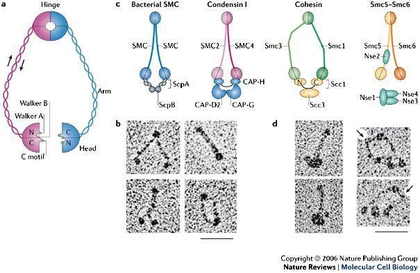
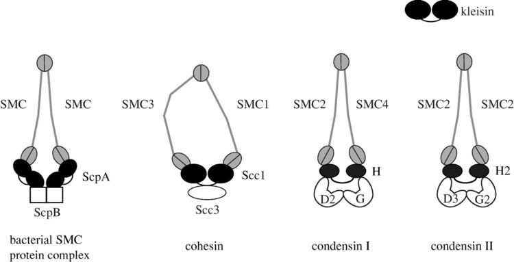
Eukaryotes have at least six SMC proteins in individual organisms, and they form three distinct heterodimers with specialized functions:
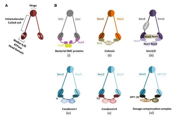
Each complex contains a distinct set of non-SMC regulatory subunits. Some organisms have variants of SMC proteins. For instance, mammals have a meiosis-specific variant of SMC1, known as SMC1β. The nematode Caenorhabditis elegans has an SMC4-variant that has a specialized role in dosage compensation.
Prokaryotic SMCs
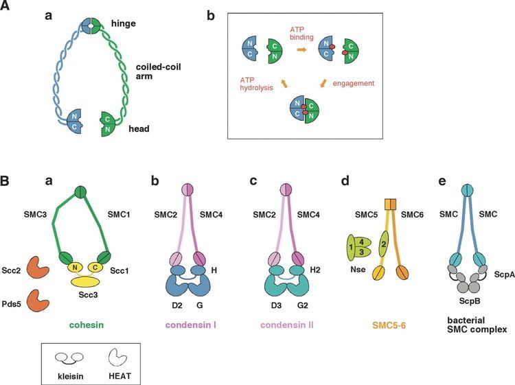
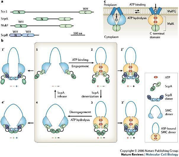
SMC proteins are conserved from bacteria to humans. Most bacteria have a single SMC protein in individual species that forms a homodimer. In a subclass of Gram-negative bacteria including Escherichia coli, a distantly related protein known as MukB plays an equivalent role.
Primary structure
SMC proteins are 1,000-1,500 amino-acid long. They have a modular structure that is composed of the following domains:
- Walker A ATP-binding motif
- coiled-coil region I
- hinge region
- coiled-coil region II
- Walker B ATP-binding motif; signature motif
Secondary and tertiary structure
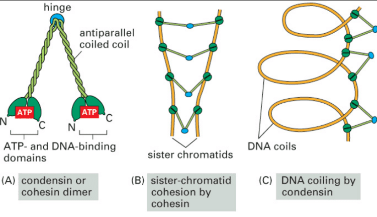
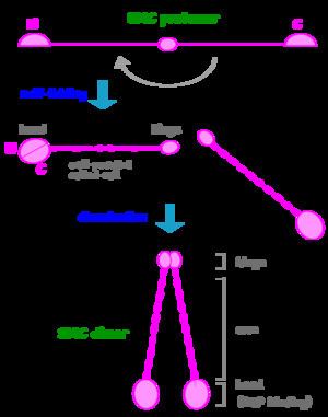
SMC dimers form a V-shaped molecule with two long coiled-coil arms. To make such a unique structure, an SMC protomer is self-folded through anti-parallel coiled-coil interactions, forming a rod-shaped molecule. At one end of the molecule, the N-terminal and C-terminal domains together form an ATP-binding domain. The other end is called a hinge domain. Two protomers then dimerize through their hinge domains and assemble a V-shaped dimer. The length of the coiled-coil arms is ~50 nm long. Such long "antiparallel" coiled-coils are very rare, and found only among SMC proteins (and its relatives such as Rad50). The ATP-binding domain of SMC proteins is structurally related to that of ABC transporters, a large family of transmembrane proteins that actively transport small molecules across cellular membranes. It is thought that the cycle of ATP binding and hydrolysis modulates the cycle of closing and opening of the V-shaped molecule, but the detailed mechanisms of action of SMC proteins remain to be determined.
Genes
The following human genes encode SMC proteins: