
HUGO 9281 RefSeq NP_002699.1 | Entrez 5499 OMIM 176875 | |
 | ||
Alt. symbols PP1, PP1a, MGC15877, MGC1674, PP-1A, PP1alpha, PPP1A | ||
Protein phosphatase 1 (PP1) belongs to a certain class of phosphatases known as protein serine/ threonine phosphatases. This type of phosphatase includes metal-dependent protein phosphatases (PPMs) and aspartate-based phosphatases. PP1 has been found to be important in the control of glycogen metabolism, muscle contraction, cell progression, neuronal activities, splicing of RNA, mitosis, cell division, apoptosis, protein synthesis, and regulation of membrane receptors and channels.
Contents
Structure
Each PP1 enzyme contains both a catalytic subunit and a regulatory subunit. The catalytic subunit consists of a 30-kD single-domain protein that can form complexes with other regulatory subunits. The catalytic subunit is highly conserved among all eukaryotes, thus suggesting a common catalytic mechanism. The catalytic subunit can from complexes with various regulatory subunits. These regulatory subunits play an important role in substrate specificity as well as compartmentalization. Some common regulatory subunits include GM and GL, which are named after their locations of action within the body (Muscle and Liver respectively).
X-ray crystallographic structural data is available for PP1 catalytic subunit. The catalytic subunit of PP1 forms an α/β fold with a central β-sandwich arranged between two α-helical domains. The interaction of the three β-sheets of the β-sandwich creates a channel for catalytic activity, as it is the site of coordination of metal ions. These metal ions have been identified as Mn and Fe and their coordination is provided by three histidines, two aspartic acids, and one asparagine.
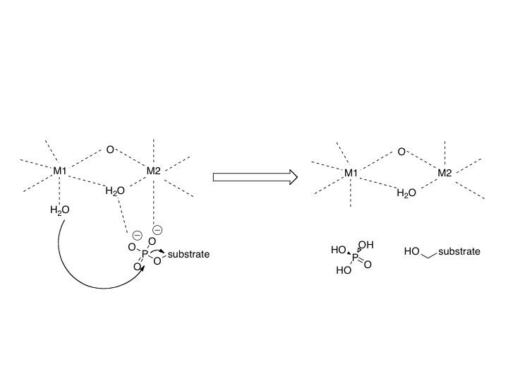
Mechanism
The mechanism involves two metal ions binding and activating water, which initiates a nucleophilic attack on the phosphorus atom.
Regulation
Regulation of these different processes is performed by distinct PP1 holoenzymes that facilitate the complexation of the PP1 catalytic subunit to various regulatory subunits.
Potential inhibitors include a variety of naturally occurring toxins including okadaic acid, a diarrhetic shellfish poison, strong tumor promoter, and microcystin. Microcystin is a liver toxin produced by blue-green algae and contains a cyclic heptapeptide structure that interacts with three distinct regions of the surface of the catalytic subunit of PP1. The structure of MCLR does not change when compexed with PP1, but the catalytic subunit of PP1 does in order to avoid steric effects of Tyr 276 of PP1 and Mdha side chain of MCLR.
Biological function
PP1 plays a crucial role in the regulation of blood-glucose levels in the liver and glycogen metabolism. PP1 is important to the reciprocal regulation of glycogen metabolism by ensuring the opposite regulation of glycogen breakdown and glycogen synthesis.
Phosphorylase a serves as a glucose sensor in liver cells. When glucose levels are low, phosphorylase a in its active R state has PP1 bound tightly. This binding to phosphorylase a prevents any phosphatase activity of PP1 and maintains the active phosphorylated configuration. Therefore, there phosphorylase a will accelerate glycogen breakdown until adequate levels of glucose are achieved. When glucose concentrations get too high, phosphorylase a is converted to its inactive, T state. By shifting phosphorylase a to its T state, PP1 dissociates from the complex. This dissociation activates glycogen synthase and converts phosphorylase a to phosphorylase b. It should be noted that phosphorylase b does not bind PP1 allowing PP1 to remain activated.
When the muscles of the body signal for the need for glycogen degradation and increased glucose concentration, PP1 will be regulated accordingly. Protein kinase A can reduce the activity of PP1. The glycogen binding region, GM, becomes phosphorylated, which causes its dissociation from the catalytic PP1 unit. This separation of the catalytic PP1 unit, glycogen, and other substrates causes a significant decrease in dephosphorylation. Also, when other substrates become phosphorylated by protein kinase A, they can bind to the catalytic subunit of PP1 and directly inhibit it. In the end, phosphorylase is kept in its active form and glycogen synthase in its inactive form.
Disease relevance
In Alzheimer’s, hyperphosphorylation of the microtubule-associated protein inhibits the assembly of microtubules in neurons. Researchers at the New York State Institute for Basic Research in Developmental Disabilities showed that there is significantly lower type 1 phosphatase activity in both gray and white matters in Alzheimer disease brains. This suggests that dysfunctional phosphatases play a role in Alzheimer's disease.
Regulation of HIV-1 transcription by Protein Phosphatase 1 (PP1). It has been recognized that protein phosphatase-1 (PP1) serves as an important regulator of HIV-1 transcription. Researchers at Howard University showed that Tat protein targets PP1 to the nucleus and the consequent interaction is important for HIV-1 transcription. The protein also contributes to ebolavirus pathogenesis by dephosphorylating the viral transcription activator VP30, allowing it to produce viral mRNAs. Inhibition of PP1 prevents VP30 dephosphorylation, thus preventing manufacture of viral mRNA, and thus viral protein. The viral L polymerase is, however, still capable of replicating viral genomes without VP30 dephosphorylation by PP1.
The herpes simplex virus protein ICP34.5 also activates protein phosphatase 1, which overcomes the cellular stress response to viral infection; protein kinase R is activated by the virus' double-stranded RNA, and protein kinase R then phosphorylates a protein called eukaryotic initiation factor-2A (eIF-2A), which inactivates eIF-2A. EIF-2A is required for translation so by shutting down eIF-2A, the cell prevents the virus from hijacking its own protein-making machinery. Herpesviruses in turn evolved ICP34.5 to defeat the defense; ICP34.5 activates protein phosphatase-1A which dephosphorylates eIF-2A, allowing translation to occur again.
Subunits
Protein phosphatase 1 is a multimeric enzyme that contains the following subunits: