
Symbol PROZ HUGO 9460 PDB 1LP1 | Entrez 8858 OMIM 176895 RefSeq NM_003891 | |
 | ||
Protein Z (PZ or PROZ) is a protein which in humans is encoded by the PROZ gene.
Contents
Protein Z is a member of the coagulation cascade, the group of blood proteins that leads to the formation of blood clots. It is a gla domain protein and thus vitamin K-dependent, and its functionality is therefore impaired in warfarin therapy. It is a glycoprotein.
Physiology
Although it is not enzymatically active, it is structurally related to several serine proteases of the coagulation cascade: factors VII, IX, X and protein C. The carboxyglutamate residues (which require vitamin K) bind protein Z to phospholipid surfaces.
The main role of protein Z appears to be the degradation of factor Xa. This is done by protein Z-related protease inhibitor (ZPI), but the reaction is accelerated 1000-fold by the presence of protein Z. Oddly, ZPI also degrades factor XI, but this reaction does not require the presence of protein Z.
In some studies, deficiency states have been associated with a propensity to thrombosis. Others, however, link it to bleeding tendency; there is no clear explanation for this, as it acts physiologically as an inhibitor, and deficiency would logically have led to a predisposition for thrombosis.
Genetics
It is 62 kDa large and 396 amino acids long. The PROZ gene has been linked to the thirteenth chromosome (13q34).
It has four domains: a gla-rich region, two EGF-like domains and a trypsin-like domain. It lacks the serine residue that would make it catalytically active as a serine protease.
History
Protein Z was first isolated in cattle blood by Prowse and Esnouf in 1977, and Broze & Miletich determined it in human plasma in 1984.
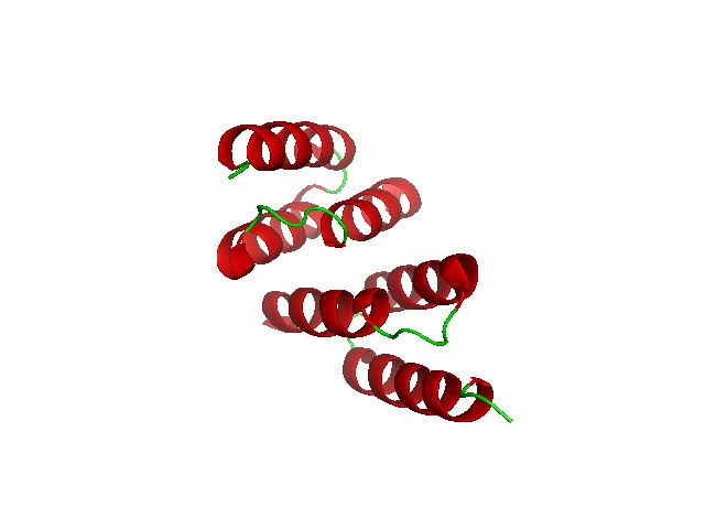
Structure
Structural analysis of protein Z will allow better understanding of its function. The Ramachandran plot for protein Z indicates it will form alpha helices. The final structure, all alpha domain, was determined by x-ray diffraction. It consists of chain A and B, which are both helix-loop-helix motifs.