
MeSH A08.800.800.120.910 FMA 50869 | TA A14.2.01.121 | |
 | ||
Latin Nervus vestibulocochlearis | ||
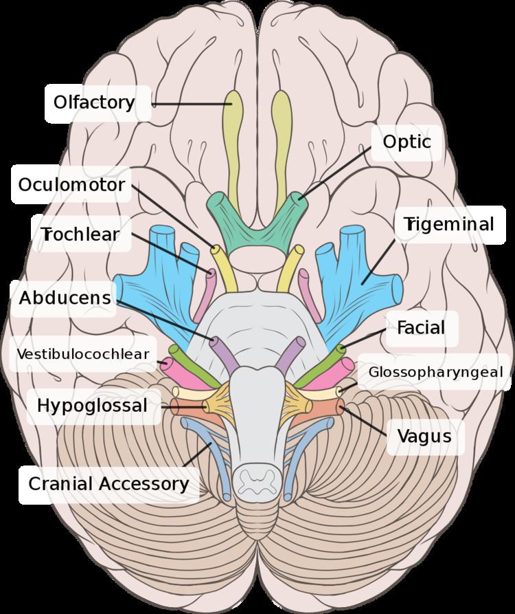
The vestibulocochlear nerve (auditory vestibular nerve), known as the eighth cranial nerve, transmits sound and equilibrium (balance) information from the inner ear to the brain.
Contents
Structure
The vestibulocochlear nerve consists mostly of bipolar neurons and splits into two large divisions: the cochlear nerve and the vestibular nerve.
The cochlear nerve travels away from the cochlea of the inner ear where it starts as the spiral ganglia. Processes from the organ of Corti conduct afferent transmission to the spiral ganglia. It is the inner hair cells of the organ of Corti that are responsible for activation of afferent receptors in response to pressure waves reaching the basilar membrane through the transduction of sound. The exact mechanism by which sound is transmitted by the neurons of the cochlear nerve is uncertain; the two competing theories are place theory and temporal theory.
The vestibular nerve travels from the vestibular system of the inner ear. The vestibular ganglion houses the cell bodies of the bipolar neurons and extends processes to five sensory organs. Three of these are the cristae located in the ampullae of the semicircular canals. Hair cells of the cristae activate afferent receptors in response to rotational acceleration. The other two sensory organs supplied by the vestibular neurons are the maculae of the saccule and utricle. Hair cells of the maculae in the utricle activate afferent receptors in response to linear acceleration while hair cells of the maculae in the saccule respond to vertically directed linear force.
Development
The vestibulocochlear nerve is derived from the embryonic otic placode.
Function
This is the nerve along which the sensory cells (the hair cells) of the inner ear transmit information to the brain. It consists of the cochlear nerve, carrying information about hearing, and the vestibular nerve, carrying information about balance. It emerges from the pontomedullary junction and exits the inner skull via the internal acoustic meatus (or internal auditory meatus) in the temporal bone.
The vestibulocochlear nerve carries axons of type SSA, special somatic afferent, which carry the modalities of hearing and equilibrium.
Symptoms of damage
Damage to the vestibulocochlear nerve may cause the following symptoms:
Examination
Examinations that can be done include the Rinnes' test and the Weber Test
Etymology
Some older texts call the nerve the acoustic or auditory nerve, but these terms have fallen out of widespread use because they fail to recognize the nerve's role in the vestibular system. Vestibulocochlear nerve is therefore preferred by most.