
Specialty gastroenterology ICD-9-CM 568.89, 770.2 eMedicine radio/562 | ICD-10 K66.8 DiseasesDB 31511 MeSH D011027 | |
 | ||
Pneumoperitoneum is pneumatosis (abnormal presence of air or other gas) in the peritoneal cavity, a potential space within the abdominal cavity. The most common cause is a perforated abdominal viscus, generally a perforated peptic ulcer, although any part of the bowel may perforate from a benign ulcer, tumor or abdominal trauma. A perforated appendix seldom causes a pneumoperitoneum.
Contents
In the mid-twentieth century, an "artificial" pneumoperitoneum was sometimes intentionally administered as a treatment for a hiatal hernia. This was achieved by insufflating the abdomen with carbon dioxide. The practice is currently used by surgical teams in order to perform laparoscopic surgery.
Causes
Diagnosis
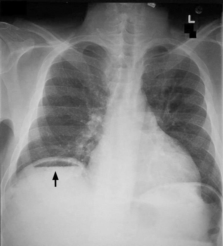
When present, pneumoperitoneum can often be seen on projectional radiography, but small amounts are often missed, and CT scan is nowadays regarded as a criterion standard in the assessment of a pneumoperitoneum. CT can visualize quantities as small as 5 cm³ of air or gas.
Signs that can be seen on projectional radiography are the double wall sign (also called Rigler's sign) and the football sign.
The double wall sign marks the presence of air on both sides of the intestine. However, a false double wall sign can result from two loops of bowel being in contact with one another. The sign is named after Leo George Rigler. It is not the same as Rigler's triad.
The football sign is when the abdomen appears as a large oval radiolucency reminiscent of an American football on a supine projectional radiograph. The football sign is most frequently seen in infants with spontaneous or iatrogenic gastric perforation causing pneumoperitoneum. It is also seen in bowel obstruction with secondary perforation, as in Hirschprung disease, midgut volvulus, meconium ileus and intestinal atresia. Iatrogenic causes like endoscopic perforation may also give football sign.
As differential diagnoses, a subphrenic abscess, bowel interposed between diaphragm and liver (Chilaiditi syndrome), and linear atelectasis at the base of the lungs can simulate free air under the diaphragm on a chest X-ray.
Terminology
Pneumoperitoneum can be described as peritoneal emphysema, just as pneumomediastinum can be called mediastinal emphysema, but pneumoperitoneum is the usual name.