
ICD-10-PCS B?0 OPS-301 code 3-10...3-13 | ICD-9-CM 87 | |
 | ||
Projectional radiography is the practice of producing two-dimensional images using x-ray radiation. Radiographic exams are typically performed by radiographers, trained, licensed medical professionals. Projectional radiography is the cornerstone of modern medical imaging, and can be used to image almost every part of the human body. Mammography, DXA and dental radiography are specialized variants of projectional radiography. Computed tomography also uses x-radiation, but its different image acquisition parameters allow for images to be acquired in the axial plane.
Contents
Imaging principles
Projectional radiography relies on the characteristics of x-ray radiation (quantity and quality of the beam) and knowledge of how it interacts with human tissue to create diagnostic images. X-rays are a form of ionizing radiation, meaning it has sufficient energy to potentially remove electrons from an atom, thus giving it a charge and making it an ion. Ionizing radiation has sufficient energy to penetrate human tissue.
X-ray attenuation
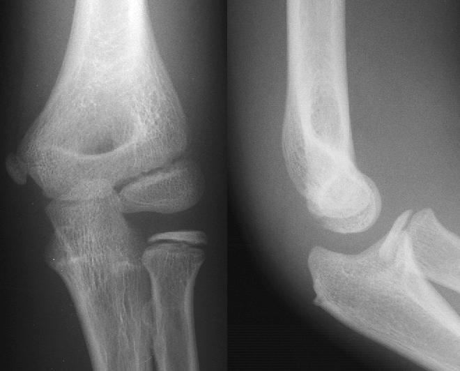
When an exposure is made, x-ray radiation exits the tube as what is known as the primary beam. When the primary beam passes through the body, some of the radiation is absorbed in a process known as attenuation. Anatomy that is denser has a higher rate of attenuation than anatomy that is less dense, so bone will absorb more x-rays than soft tissue. What remains of the primary beam after attenuation is known as the remnant beam. The remnant beam is responsible for exposing the image receptor. Areas on the image receptor that receive the most radiation (portions of the remnant beam experiencing the least attenuation) will be more heavily exposed, and therefore will be processed as being darker. Conversely, areas on the image receptor that receive the least radiation (portions of the remnant beam experience the most attenuation) will be less exposed and will be processed as being lighter. This is why bone, which is very dense, process as being ‘white’ on radio graphs, and the lungs, which contain mostly air and is the least dense, shows up as ‘black’.
Density
Radiographic density is the measure of overall darkening of the image. Density is a logarithmic unit that describes the ratio between light hitting the film and light being transmitted through the film. A higher radiographic density represents more opaque areas of the film, and lower density more transparent areas of the film.
With digital imaging, however, density may be referred to as brightness. The brightness of the radiograph in digital imaging is determined by computer software and the monitor on which the image is being viewed.
Contrast
Contrast is defined as the difference in radiographic density between adjacent portions of the image. The range between black and white on the final radiograph. High contrast, or short-scale contrast, means there is little gray on the radiograph, and there are fewer gray shades between black and white. Low contrast, or long-scale contrast, means there is much gray on the radiograph, and there are many gray shades between black and white.
Closely related to radiographic contrast is the concept of exposure latitude. Exposure latitude is the range of exposures over which the recording medium (image receptor) will respond with a diagnostically useful density; in other words, this is the "flexibility" or "leeway" that a radiographer has when setting his/her exposure factors. Images having a short-scale of contrast will have narrow exposure latitude. Images having long-scale contrast will have a wide exposure latitude; that is, the radiographer will be able to utilize a broader range of technical factors to produce a diagnostic-quality image.
Contrast is determined by the kilovoltage (kV; energy/quality/penetrability) of the x-ray beam and the tissue composition of the body part being radiographed. Selection of look-up tables (LUT) in digital imaging also affects contrast.
Generally speaking, high contrast is necessary for body parts in which bony anatomy is of clinical interest (extremities, bony thorax, etc). When soft tissue is of interest (ex. abdomen or chest), lower contrast is preferable in order to accurately demonstrate all of the soft tissue tones in these areas.
Variations by target organs
Projection radiography uses X-rays in different amounts and strengths depending on what body part is being imaged:
Projectional radiography terminology
NOTE: The word 'view' is often used erroneously to describe a radiographic projection.
Equipment Used in Projectional Radiography
Routine projections used in the US
Chest - (CXR) Includes a PA and Lateral with the patient standing or sitting up. Special projections include an AP in cases where the image needs to be obtained stat and with a portable device, particularly when a patient cannot be safely positioned upright. Lateral Decubitus may be used for visualization of air-fluid levels if an upright image cannot be obtained. AP Axial Lordotic projects the clavicles above the lung fields, allowing better visualization of the apices (which is extremely useful when looking for evidence of primary tuberculosis)
Abdomen - Usually a single AP supine (KUB—kidney, bladder, and ureter) projection. Special projections include a PA prone, Lateral Decubitus, upright AP, and Lateral Cross-Table (with the patient supine) A minimal acute obstructive series (for the purpose of ruling out small bowel obstruction) would include two views: typically, a supine view and an upright view (which would be sufficient to detect air-fluid levels), although a lateral decubitus could be substituted for the upright.
Cervical Spine - Five or six projections are common; a Lateral, two 45 degree obliques, an AP axial (Cephalad), an AP "Open Mouth" for C1-C2, and Cervicothoracic Lateral (Swimmer's) to better visualize C7-T1 if necessary. Special projections include a Lateral with Flexion and Extension of the cervical spine, an Axial for C1-C2 (Fuchs or Judd method), and an AP Axial (Caudad) for articular pillars.
Thoracic Spine - An AP and Lateral are basic projections. Obliques 20 degrees from lateral may be ordered to better visualize the zygapophysial joint
Lumbar Spine - Basic projections include an AP, two Obliques, a Lateral, and a Lateral L5-S1 spot to better visualize the L5-S1 interspace. Special projections are AP Right and Left bending, and Laterals with Flexion and Extension.
Sacrum and Coccyx - If both bones are to be examined separate cephalad and caudad AP axial projections are obtained for the sacrum and coccyx respectively as well as a single Lateral of both bones.
Sternum - The two basic projections are a 15 to 20 degree Right Anterior Oblique and a Lateral.
Sternoclavicular Joints - Are usually ordered as a single PA and a Right and Left 15 degree Right Anterior Obliques.
Ribs - Common rib projections are based on the location of the area of interest. These are obtained with shorter wavelengths/higher frequencies/higher levels of radiation than a standard CXR.