
Specialty medical genetics ICD-9-CM 751.1 751.2 DiseasesDB 31514 33000 | ICD-10 Q41, Q42 OMIM 223400 243600 | |
 | ||
Intestinal atresia is a malformation where there is a narrowing or absence of a portion of the intestine. This defect can either occur in the small or large intestine.
Contents
Types
The different types of intestinal atresia are named after their location:
Duodenal atresia has a strong association with Down syndrome. It is the most common type, followed by ileal atresia.
An inherited form – familial multiple intestinal atresia – has also been described. This disorder was first reported in 1971. It is due to a mutation in the gene TTC7A on short arm of chromosome 2 (2p16). It is inherited as an autosomal recessive gene and is usually fatal in infancy.
Cause
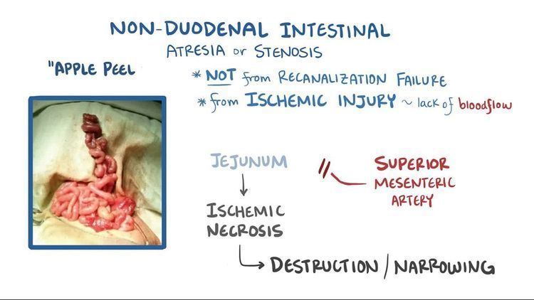
The most common cause of non-duodenal intestinal atresia is a vascular accident in utero that leads to decreased intestinal perfusion and ischemia of the respective segment of bowel. This leads to narrowing, or in the most severe cases, complete obliteration of the intestinal lumen.
In the case that the superior mesenteric artery, or another major intestinal artery, is occluded, large segments of bowel can be entirely underdeveloped. Classically, the affected area of bowel assumes a spiral configuration and is described to have an "apple peel" like appearance; this is accompanied by lack of a dorsal mesentery. Ileal atresia can also result as a complication of meconium ileus.
Diagnosis
Intestinal atresias are often discovered before birth: either during a routine sonogram which shows a dilated intestinal segment due to the blockage, or by the development of polyhydramnios (the buildup of too much amniotic fluid in the uterus). These abnormalities are indications that the fetus may have a bowel obstruction which a more detailed ultrasound study can confirm.
Some fetuses with bowel obstruction have abnormal chromosomes. An amniocentesis is recommended because it can determine not only the sex of the baby, but whether or not there is a problem with the chromosomes.
Treatment
Fetal and neonatal intestinal atresia are treated using laparotomy after birth. If the area affected is small, the surgeon may be able to remove the damaged portion and join the intestine back together. In instances where the narrowing is longer, or the area is damaged and cannot be used for period of time, a temporary stoma may be placed.