ICD-10-PCS [1] OPS-301 code 3-800, 3-820 | ICD-9-CM 88.91 | |
 | ||
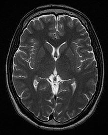
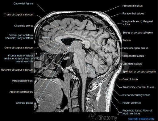
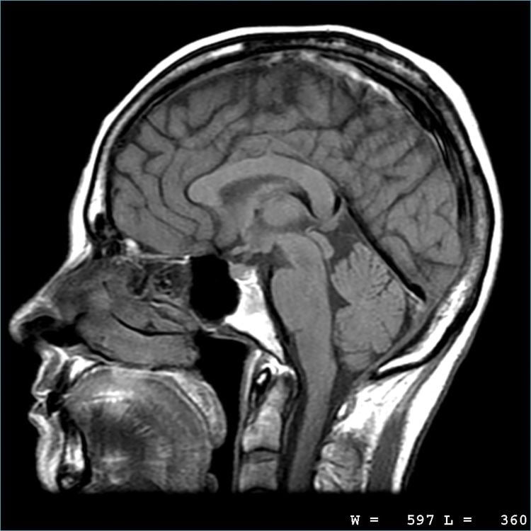
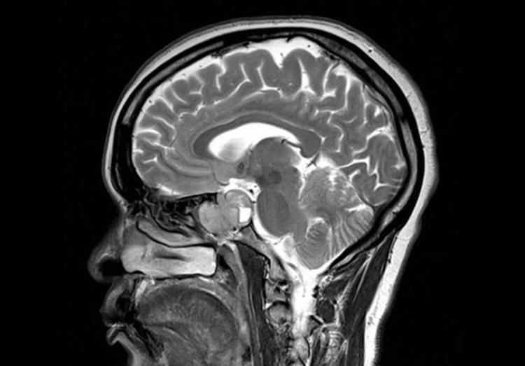
Magnetic resonance imaging (MRI) of the nervous system uses magnetic fields and radio waves to produce high quality two- or three-dimensional images of nervous system structures without use of ionizing radiation (X-rays) or radioactive tracers.
Contents
History
The first MR images of a human brain were obtained in 1978 by two groups of researchers at EMI Laboratories led by Ian Robert Young and Hugh Clow. In 1986, Charles L. Dumoulin and Howard R. Hart at General Electric developed MR angiography and Denis Le Bihan, obtained the first images and later patented diffusion MRI. In 1990, Seiji Ogawa at AT&T Bell labs recognized that oxygen-depleted blood with dHb was attracted to a magnetic field, and discovered the technique that underlies Functional Magnetic Resonance Imaging (fMRI). In 1997, Jürgen R. Reichenbach, E. Mark Haacke and coworkers at Washington University developed Susceptibility weighted imaging. The first study of the human brain at 3.0 T was published in 1994, and in 1998 at 8 T. Studies of the human brain have been performed at up to 9.4 T. Paul Lauterbur and Sir Peter Mansfield were awarded the 2003 Nobel Prize in Physiology or Medicine for their discoveries concerning MRI.
Applications
One advantage of MRI of the brain over computed tomography of the head is better tissue contrast, and it has fewer artifacts than CT when viewing the brainstem. MRI is also superior for pituitary imaging. It may however be less effective at identifying early cerebritis.
In the case of a concussion, an MRI should be avoided unless there are progressive neurological symptoms, focal neurological findings or concern of skull fracture on exam.
In analysis of the fetal brain, MRI provides more information about gyration than ultrasound.
A number of different imaging modes can be used with imaging the nervous system: