
ICD-9-CM 87.03 | OPS-301 code 3-200, 3-220 | |
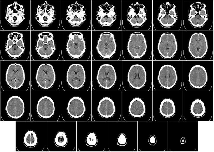 | ||
Computed tomography (CT) of the head or Computed Axial Tomography (CAT) scanning uses a series of x-rays of the head taken from many different directions. Typically used for quickly viewing brain injuries, CT scanning uses a computer program that performs a numerical integral calculation (the inverse Radon transform) on the measured x-ray series to estimate how much of an x-ray beam is absorbed in a small volume of the brain. Typically the information is presented as a series of cross sections of the brain.
Contents
In approximation, the denser a material is, the whiter a volume of it will appear on the scan, just as in the more familiar "flat" X-rays, where dense bone appears white. CT scans are primarily used for evaluating swelling from tissue damage in the brain, bleeding, and in assessment of ventricle size. Modern CT scanning can provide reasonably good images in a matter of minutes. A spiral CT scan of the head may be performed in 10–30 seconds, making it a good option for children and adults with difficulty holding still for longer periods. The use of contrast dye in CT angiography provides good visualization of the vascular structures and leaks in the blood vessels.
Indications
Computed tomography (CT) has become the diagnostic modality of choice for head trauma due to its accuracy, reliability, safety, and wide availability. The changes in microcirculation, impaired auto-regulation, cerebral edema, and axonal injury start as soon as head injury occurs and manifest as clinical, biochemical, and radiological changes. Proper therapeutic management of brain injury is based on correct diagnosis and appreciation of the temporal course of the disease process. CT scan detects and precisely localizes the intracranial hematomas, brain contusions, edema and foreign bodies. Because of the widespread availability of CT, there is reduction in arteriography, surgical intervention and skull radiography.
Even in emergency situations, when a head injury is minor as determined by a physician's evaluation and based on established guidelines, CT of the head should be avoided for adults and delayed pending clinical observation in the emergency department for children. Many people visit emergency departments for minor head injuries. CT scans of the head can confirm a diagnosis of skull fracture or brain bleeding, but even in the emergency room such things are uncommon and not minor injuries so CT of the head is usually not necessary. Clinical trials have shown the efficacy and safety of using CT of the head in emergency settings only when indicated, which would be at the indication of evidence-based guidelines following the physical examination and a review of the patient's history.
Concussion is not a routine indication for having brain CT or brain MRI and can be diagnosed by a healthcare provider trained to manage concussions. People with concussion usually do not have relevant abnormalities about which brain imaging could give insight, and so brain imaging should not routinely be ordered for people with concussion. If there is concern about a skull fracture, focal neurological symptoms present or worsening symptoms, then CT imaging may be useful. MRI may be useful for people whose symptoms worsen over time or when structural pathology is suspected.
CT of the head is sometimes used for patients who have sudden hearing loss. This use is not indicated and offers no information which would improve the initial management of the patient. In patients for which there are not other neurological findings, a history of trauma, or a history of ear disease, CT scans should not be used in response to sudden hearing loss.
CT of the head is also used in CT-guided stereotactic surgery and radiosurgery for treatment of intracranial tumors, arteriovenous malformations and other surgically treatable conditions using a device known as the N-localizer.
Orbital views for eye-related disorders
Special views focusing on the orbit of the eye may be taken to investigate concerns relating to the eye. CT scans are used by physicians specializing in treating the eye (ophthalmologists) to detect foreign bodies (especially metallic objects), fractures, abscesses, cellulitis, sinusitis, bleeding within the skull (intracranial bleeding), proptosis, Graves disease changes in the eye, and evaluation of the orbital apex and cavernous sinus.
Comparison with MRI
Magnetic resonance imaging (MRI) of the head provides superior information as compared to CT scans when seeking information about headache to confirm a diagnosis of neoplasm, vascular disease, posterior cranial fossa lesions, cervicomedullary lesions, or intracranial pressure disorders. It also does not carry the risks of exposing the patient to ionizing radiation. CT scans may be used to diagnose headache when neuroimaging is indicated and MRI is not available, or in emergency settings when hemorrhage, stroke, or traumatic brain injury are suspected.
MRI (magnetic resonance imaging) provides more sensitivity in the evaluation of the cavernous sinus and the orbital apex.
One advantage over a brain MRI is in the evaluation of intracerebral calcifications.
Cautions
Several different views of the head are available, including axial, coronal, reformatted coronal, and reformatted sagittal images. However, coronal images require the patient hyperextend their neck, which must be avoided if any possibility of neck injury exists.