
Specialty urology ICD-9-CM 583.81 eMedicine med/1597 | ICD-10 M32.1+N08.5* MedlinePlus 000481 MeSH D008181 | |
 | ||
Lupus nephritis (also known as SLE nephritis) is an inflammation of the kidneys caused by systemic lupus erythematosus (SLE), an autoimmune disease. It is a type of glomerulonephritis in which the glomeruli become inflamed. As the result of SLE, the cause of glomerulonephritis is said to be secondary and has a different pattern and outcome from conditions with a primary cause originating in the kidney.
Contents
Signs and symptoms
General symptoms of lupus nephritis include
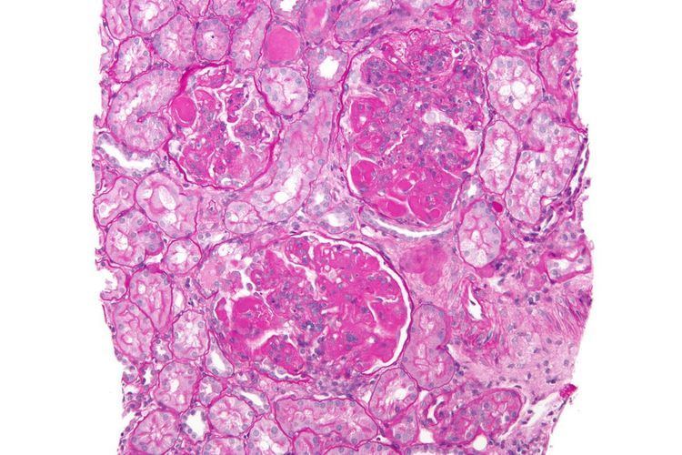
Class I disease (minimal mesangial glomerulonephritis) in its histology has a normal appearance under a light microscope, but mesangial deposits are visible under an electron microscope. At this stage urinalysis is normal.
Class II disease (mesangial proliferative glomerulonephritis) is noted by mesangial hypercellularity and matrix expansion. Microscopic haematuria with or without proteinuria may be seen. Hypertension, nephrotic syndrome, and acute kidney insufficiency are very rare at this stage.
Class III disease (focal glomerulonephritis) is indicated by sclerotic lesions involving less than 50% of the glomeruli, which can be segmental or global, and active or chronic, with endocapillary or extracapillary proliferative lesions. Under the electron microscopy, subendothelial deposits are noted, and some mesangial changes may be present. Immunofluorescence reveals positively for IgG, IgA, IgM, C3, and C1q. Clinically, haematuria and proteinuria are present, with or without nephrotic syndrome, hypertension, and elevated serum creatinine.
Class IV disease (diffuse proliferative nephritis) is both the most severe, and the most common subtype. More than 50% of glomeruli are involved. Lesions can be segmental or global, and active or chronic, with endocapillary or extracapillary proliferative lesions. Under electron microscopy, subendothelial deposits are noted, and some mesangial changes may be present. Clinically, haematuria and proteinuria are present, frequently with nephrotic syndrome, hypertension, hypocomplementemia, elevated anti-dsDNA titres and elevated serum creatinine.
Class V disease (membranous glomerulonephritis) is characterized by diffuse thickening of the glomerular capillary wall (segmentally or globally), with diffuse membrane thickening, and subepithelial deposits seen under the electron microscope. Clinically, stage V presents with signs of nephrotic syndrome. Microscopic haematuria and hypertension may also been seen. Stage V also can also lead to thrombotic complications such as renal vein thromboses or pulmonary emboli.
A final Class is included by most practitioners, Class VI, or advanced sclerosing lupus nephritis. It is represented by global sclerosis involving more than 90% of glomeruli, and represents healing of prior inflammatory injury. Active glomerulonephritis is not usually present. This stage is characterised by slowly progressive kidney dysfunction, with relatively bland urine sediment. Response to immunotherapy is usually poor. A tubuloreticular inclusion within capillary endothelial cells is also characteristic of lupus nephritis, and can be seen under an electron microscope in all stages. It is not diagnostic however, as it exists in other conditions such as HIV infection. It is thought to be due to the chronic interferon exposure.
Cause
The cause of lupus nephritis, a genetic predisposition, plays role in lupus nephritis. Multiple genes, many of which are not yet identified, mediate this genetic predisposition.
The immune system protects the human body from infection, with immune system problems it cannot distinguish between harmful and healthy substances. Lupus nephritis affects approximately 3 out of 10,000 people.
Pathophysiology
The pathophysiology of lupus nephritis has autoimmunity contributing significantly. Autoantibodies direct themselves against nuclear elements. The characteristics of nephritogenic autoantibodies ( lupus nephritis) are: antigen specificity directed at nucleosome, high affinity autoantibodies form intravascular immune complexes, autoantibodies of certain isotypes activate complement.
Diagnosis
The diagnosis of lupus nephritis depends on blood tests, urinalysis, X-rays, ultrasound scans of the kidneys, and a kidney biopsy. On urinalysis, a nephritic picture is found and red blood cell casts, red blood cells and proteinuria is found. The World Health Organization has divided lupus nephritis into five stages based on the biopsy. This classification was defined in 1982 and revised in 1995.
Treatment
Drug regimens prescribed for lupus nephritis include mycophenolate mofetil (MMF), intravenous cyclophosphamide with corticosteroids, and the immune suppressant azathioprine with corticosteroids. MMF and cyclophosphamide with corticosteroids are equally effective in achieving remission of the disease. MMF is safer than cyclophosphamide with corticosteroids, with less chance of causing ovarian failure, immune problems or hair loss. It also works better than azathioprine with corticosteroids for maintenance therapy. Individuals with lupus nephritis have a high risk for B-cell lymphoma (which begins in the immune system cells).