
EC number 1.21.99.4 ExPASy NiceZyme view | CAS number 70712-46-8 | |
 | ||
Iodothyronine deiodinases (EC 1.21.99.4 and EC 1.21.99.3) are a subfamily of deiodinase enzymes important in the activation and deactivation of thyroid hormones. Thyroxine (T4), the precursor of 3,5,3'-triiodothyronine (T3) is transformed into T3 by deiodinase activity. T3, through binding a nuclear thyroid hormone receptor, influences the expression of genes in practically every vertebrate cell. Iodothyronine deiodinases are unusual in that these enzymes contain selenium, in the form of an otherwise rare amino acid selenocysteine.
Contents
- Activation and inactivation
- Structure
- Types
- Function
- Disease relevance
- Quantifying enzyme activity
- References
These enzymes are not to be confused with the iodotyrosine deiodinases that are also deiodinases, but not members of the iodothyronine family. The iodotyrosine deiodinases (unlike the iodothyronine deiodinases) do not use selenocysteine or selenium. The iodotyrosine enzymes work on iodinated single tyrosine residue molecules to scavenge iodine, and do not use as substrates the double-tyrosine residue molecules of the various iodothyronines.
Activation and inactivation
In tissues, deiodinases can either activate or inactivate thyroid hormones:
The major part of thyroxine deiodination occurs within the cells.
Deionidase 2 activity can be regulated by ubiquitination:
D-propranolol inhibits thyroxine deiodinase, thereby blocking the conversion of T4 to T3, providing some though minimal therapeutic effect.
Structure
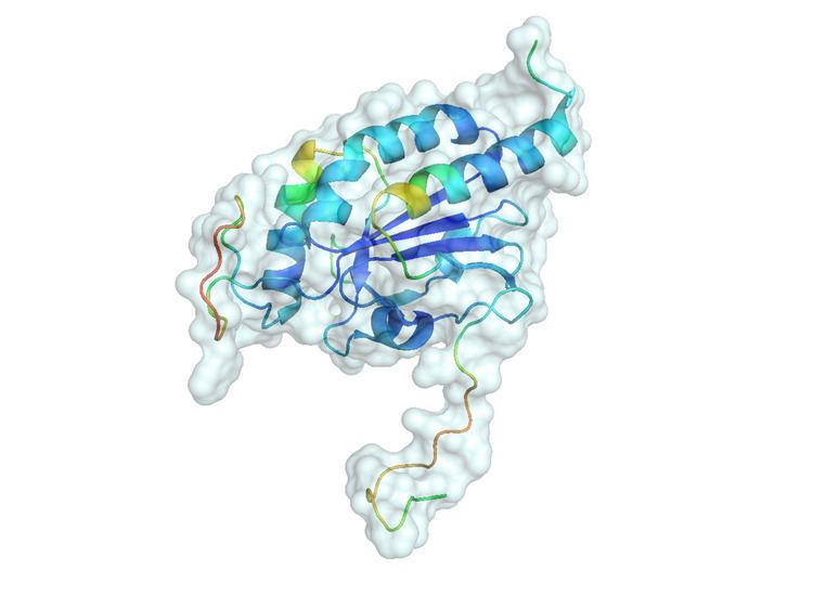
The three deiodinase enzymes share certain structural features in common although their sequence identity is lower than 50%. Each enzyme weighs between 29 and 33kDa. Deiodinases are dimeric integral membrane proteins with single transmembrane segments and large globular heads (see below). They share a TRX fold that contains the active site including the rare selenocysteine amino acid and two histidine residues. Selenocysteine is coded by a UGA codon, which generally signifies termination of a peptide through a stop codon. In point mutation experiments with Deiodinase 1 changing UGA to the stop codon TAA resulted in a complete loss of function, while changing UGA to cysteine (TGT) caused the enzyme to operate at around 10% normal efficiency. In order for UGA to be read as a selenocysteine amino acid instead of a stop codon, it is necessary that a downstream stem loop sequence, the selenocysteine insertion sequence (SECIS), be present to bind with SECIS binding protein-2 (SBP-2), which binds with elongation factor EFsec. The translation of selenocysteine is not efficient, even though it is important to the functioning of the enzyme. Deiodinase 2 is localized to the ER membrane while Deiodinase 1 and 3 are found in the plasma membrane.
The related catalytic domains of Deiodinases 1-3 feature a thioredoxine-related peroxiredoxin fold. The enzymes catalyze a reductive elimination of iodine, thereby oxidizing themselves similar to Prx, followed by a reductive recycling of the enzyme.
Types
In most vertebrates, there are three types of enzymes that can deiodinate thyroid hormones:
Function
Deiodinase 1 both activates T4 to produce T3 and inactivates T4. Besides its increased function in producing extrathyroid T3 in patients with hyperthyroidism, its function is less well understood than D2 or D3 Deiodinase 2, located in the ER membrane, converts T4 into T3 and is a major source of the cytoplasmic T3 pool. Deiodinase 3 prevents T4 activation and inactivates T3. D2 and D3 are important in homeostatic regulation in maintaining T3 levels at the plasma and cellular levels. In hyperthyroidism D2 is down regulated and D3 is upregulated to clear extra T3, while in hypothyroidism D2 is upregulated and D3 is downregulated to increase cytoplasmic T3 levels.
Serum T3 levels remain fairly constant in healthy individuals, but D2 and D3 can regulate tissue specific intracellular levels of T3 to maintain homeostasis since T3 and T4 levels may vary by organ. Deiodinases also provide spatial and temporal developmental control of thyroid hormone levels. D3 levels are highest early in development and decrease over time, while D2 levels are high at moments of significant metamorphic change in tissues. Thus D2 enables production of sufficient T3 at necessary time points while D3 may shield tissue from overexposure to T3.
Deiodinase 2 also plays a significant role in thermogenesis in brown adipose tissue (BAT). In response to sympathetic stimulation, dropping temperature, or overfeeding BAT, D2 increases oxidation of fatty acids and uncouples oxidative phosphorylation via uncoupling protein, causing mitochondrial heat production. D2 increases during cold stress in BAT and increases intracellular T3 levels. In D2 deficient models, shivering is a behavioral adaptation to the cold. However, heat production is much less efficient than uncoupling lipid oxidation.
Disease relevance
In cardiomyopathy the heart reverts to a fetal gene programming due to the overload of the heart. Like during fetal development, thyroid hormone levels are low in the overloaded heart tissue in a local hypothyroid state, with low levels of deiodinase 1 and deiodinase 2. Although deiodinase 3 levels in a normal heart are generally low, in cardiomyopathy deiodinase 3 activity is increased to decrease energy turnover and oxygen consumption.
Hypothyroidism is a disease diagnosed by decreased levels of serum thyroxine (T4). Presentation in adults leads to decreased metabolism, increased weigh gain, and neuropsychiatric complications. During development, hypothyroidism is considered more severe and leads to neurotoxicity as cretinism or other human cognitive disorders, altered metabolism and underdeveloped organs. Medication and environmental exposures can result in hypothyroidism with changes in deiodinase enzyme activity. The drug iopanoic acid (IOP) decreased cutaneous cell proliferation by inhibition of deiodinase enzyme type 1 or 2 reducing the conversion of T4 to T3. The environmental chemical DE-71, a PBDE pentaBDE brominated flame retardant decreased hepatic deiodinase I transcription and enzyme activity in neonatal rats with hypothyroidism.
Quantifying enzyme activity
In vitro, including cell culture experiments, deiodination activity is determined by incubating cells or homogenates with high amounts of labeled thyroxine (T4) and required cosubstrates. As a measure of deiodination, the production of radioactive iodine and other physiological metabolites, in particular T3 or reverse T3, are determined and expressed (e.g. as fmol/mg protein/minute).
In vivo, deiodination activity is estimated from equilibrium levels of free T3 and free T4. A simple approximation is T3/T4 ratio, a more elaborate approach is calculating sum activity of peripheral deiodinases (GD) from free T4, free T3 and parameters for protein binding, dissociation and hormone kinetics.