
Latin textus adiposus fuscus FMA 20118 | TH H2.00.04.0.0004 | |
 | ||
Brown adipose tissue (BAT) or brown fat makes up the adipose organ together with white adipose tissue (or white fat). BAT is found in almost all mammals.
Contents
Classification of brown fat refers to two distinct cell populations with similar functions. The first shares a common embryological origin with muscle cells, found in larger "classic" deposits. The second develops from white adipocytes that are stimulated by the sympathetic nervous system. These adipocytes are found interspersed in white adipose tissue and are also named 'beige' or 'brite' .
BAT is especially abundant in newborns and in hibernating mammals. It is also present and metabolically active in adult humans, but its prevalence decreases as humans age. Its primary function is thermoregulation. In addition to heat produced by shivering muscle, BAT produces heat by non-shivering thermogenesis.
In contrast to white adipocytes, which contain a single lipid droplet, brown adipocytes contain numerous smaller droplets and a much higher number of (iron-containing) mitochondria, which gives BAT its brown appearance. Brown fat also contains more capillaries than white fat, to supply the tissue with oxygen and nutrients and distribute the produced heat throughout the body.
Anatomical location and histology
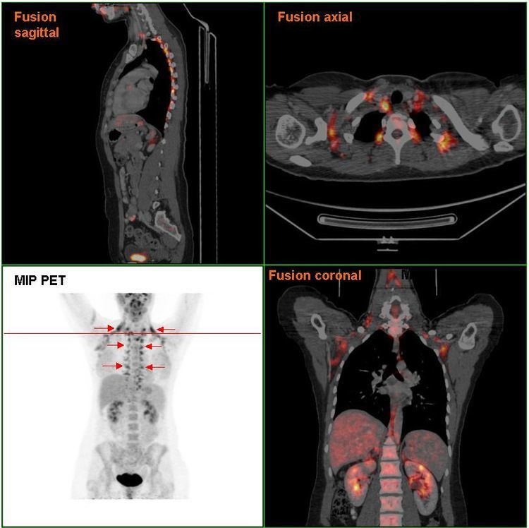
The presence of BAT in adult humans was discovered during FDG-PET scans to detect metastatic cancers. Using these scans and data from human autopsies, several BAT depots have been identified. In infants, BAT depots include, but are not limited to: interscapular, supraclavicular, suprarenal, pericardial, para-aortic and around the pancreas, kidney and trachea. These depots gradually get more white fat-like during adulthood. In adults, the depots that are most often detected in FDG-PET scans are the supraclavicular, paravertebral, mediastinal, para-aortic and suprarenal ones. It remains to be determined whether these depots are 'classical' BAT or beige/brite fat.
Brown fat in humans in the scientific and popular literature refers to two cell populations defined by both anatomical location and cellular morphology. Both share presence of small lipid droplets and numerous iron-rich mitochondria, giving the brown appearance.
Development
Brown fat cells come from the middle embryo layer, mesoderm, also the source of myocytes (muscle cells), adipocytes, and chondrocytes (cartilage cells).
The classic population of brown fat cells and muscle cells both seem to be derived from the same population of stem cells in the mesoderm, paraxial mesoderm. Both have the intrinsic capacity to activate the myogenic factor 5 (Myf5) promoter, a trait only associated with myocytes and this population of brown fat. Progenitors of traditional white fat cells and adrenergically induced brown fat do not have the capacity to activate the Myf5 promoter. Both adipocytes and brown adipocyte may be derived from pericytes, the cells which surround the blood vessels that run through white fat tissue. Notably, this is not the same as the presence of Myf5 protein, which is involved in the development of many tissues.
Additionally, muscle cells that were cultured with the transcription factor PRDM16 were converted into brown fat cells, and brown fat cells without PRDM16 were converted into muscle cells.
Function
The mitochondria in a eukaryotic cell utilize fuels to produce energy in the form of adenosine triphosphate (ATP). This process involves storing energy as a proton gradient, also known as the proton motive force (PMF), across the mitochondrial inner membrane. This energy is used to synthesize ATP when the protons flow across the membrane (down their concentration gradient) through the ATP synthase enzyme; this is known as chemiosmosis.
In endotherms, body heat is maintained by signaling the mitochondria to allow protons to run back along the gradient without producing ATP (proton leak). This can occur since an alternative return route for the protons exists through an uncoupling protein in the inner membrane. This protein, known as uncoupling protein 1 (thermogenin), facilitates the return of the protons after they have been actively pumped out of the mitochondria by the electron transport chain. This alternative route for protons uncouples oxidative phosphorylation and the energy in the PMF is instead released as heat.
To some degree, all cells of endotherms give off heat, especially when body temperature is below a regulatory threshold. However, brown adipose tissue is highly specialized for this non-shivering thermogenesis. First, each cell has a higher number of mitochondria compared to more typical cells. Second, these mitochondria have a higher-than-normal concentration of thermogenin in the inner membrane.
Infants
In neonates (newborn infants), brown fat makes up about 5% of the body mass and is located on the back, along the upper half of the spine and toward the shoulders. It is of great importance to avoid hypothermia, as lethal cold is a major death risk for premature neonates. Numerous factors make infants more susceptible to cold than adults:
Heat production in brown fat provides an infant with an alternative means of heat regulation.
Adults
It was believed that after infants grow up, most of the mitochondria (which are responsible for the brown color) in brown adipose tissue disappear, and the tissue becomes similar in function and appearance to white fat. In rare cases, brown fat continues to grow, rather than involuting; this leads to a tumour known as a hibernoma. More recent research has shown that brown fat is related not to white fat, but to skeletal muscle.
Studies using positron emission tomography scanning of adult humans have shown that BAT is still present in most adults in the upper chest and neck (especially paravertebrally). The remaining deposits become more visible (increasing tracer uptake, that is, more metabolically active) with cold exposure, and less visible if an adrenergic beta blocker is given before the scan. These discoveries could lead to new methods of weight loss, since brown fat takes calories from normal fat and burns it. Scientists have been able to stimulate brown fat growth in mice. One study of APOE knock out mice showed cold exposure could promote atherosclerotic plaque growth and instability when study mice were subjected to sustained low temperatures of 4 °C for 8 weeks, which may cause a stress condition that shows rapid forced change rather than a safe acclimatisation that can be used to understand the potential in adult humans during modest reductions of ambient temperature by just 5 to 10 °C. Furthermore, several newer studies have documented the substantial benefits of cold exposure in multiple species including humans, for example researchers concluded that "activation of BAT is a powerful therapeutic avenue to ameliorate hyperlipidaemia and protect from atherosclerosis" and that brown fat activation reduces plasma triglyceride and cholesterol levels and attenuates diet-induced atherosclerosis development.
Long term studies of adult humans are needed to establish a balance of benefit and risk, in combination with historical research of living conditions of recent human generations prior to the current increase of poor health related to excessive accumulation of white fat. Pharmacological approaches using β3-adrenoceptor agonists have been shown to enhance glucose metabolic activity of brown adipose tissue in rodents.
Additionally research has shown:
Other animals
The interscapular brown adipose tissue is commonly and inappropriately referred to as the hibernating gland. Whilst believed by many to be a type of gland, it is actually a collection of adipose tissues lying between the scapulae of rodentine mammals. Composed of brown adipose tissue and divided into two lobes, it resembles a primitive gland, regulating the output of a variety of hormones. The function of the tissue appears to be involved in the storage of medium to small lipid chains for consumption during hibernation. The smaller lipid structure allowing for a more rapid path of energy production than glycolysis.
In studies where the interscapular brown adipose tissue of rats were lesioned, it was demonstrated that the rats had difficulty regulating their normal body-weight.
The longest-lived small mammals: grey squirrels (24 yrs), bats (30 yrs), and naked mole rats (32 yrs), all have remarkably high levels of BAT and BAT activity. Furthermore, in animal species that span a wide range of latitudes, the within-species longevity is correlated with how far north (or south, in the southern hemisphere) an individual lives suggesting that cooler environments lead to increased BAT activation and increased lifespan across a wide range of species.