
 | ||
Intraocular pressure (IOP) is the fluid pressure inside the eye. Tonometry is the method eye care professionals use to determine this. IOP is an important aspect in the evaluation of patients at risk from glaucoma. Most tonometers are calibrated to measure pressure in millimeters of mercury (mmHg).
Contents
Physiology
Intraocular pressure is mainly determined by the coupling of the production of aqueous humor and the drainage of aqueous humor mainly through the trabecular meshwork located in the anterior chamber angle. The reason for this is because the vitreous humour in the posterior segment has a relatively fixed volume and thus does not affect intraocular pressure regulation.
An important quantitative relationship is provided below:
IOP = F / C + PVWhere F = aqueous fluid formation rate, C = outflow rate, PV = episcleral venous pressure. The above factors are those that drive IOP.
Measurement
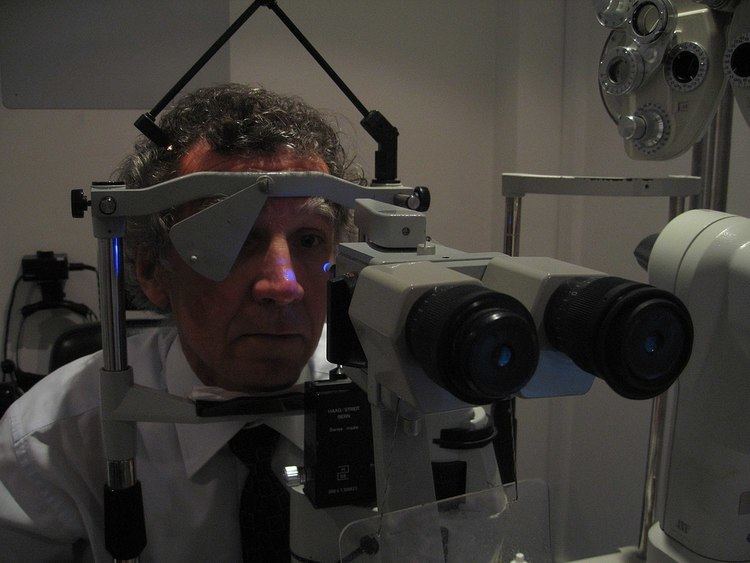
Intraocular pressure is measured with a tonometer as part of a comprehensive eye examination.
Measured values of intraocular pressure are influenced by corneal thickness and rigidity. As a result, some forms of refractive surgery (such as photorefractive keratectomy) can cause traditional intraocular pressure measurements to appear normal when in fact the pressure may be abnormally high. A newer transpalpebral and transsleral tonometry method is not influenced by corneal biomechanics and does not need to be adjusted or corneal irregularities as measurement is done over upper eyelid and sclera.
Classification
Current consensus among ophthalmologists and optometrists define normal intraocular pressure as that between 10 mmHg and 20 mmHg. The average value of intraocular pressure is 15.5 mmHg with fluctuations of about 2.75 mmHg.
Ocular hypertension (OHT) is defined by intraocular pressure being higher than normal, in the absence of optic nerve damage or visual field loss.
Hypotony, or ocular hypotony, is typically defined as intraocular pressure equal to or less than 5 mmHg. Such low intraocular pressure could indicate fluid leakage and deflation of the eyeball.
Daily variation
Intraocular pressure varies throughout the night and day. The diurnal variation for normal eyes is between 3 and 6 mmHg and the variation may increase in glaucomatous eyes. During the night, intraocular pressure may not decrease despite the slower production of aqueous humour. In the general population, IOP ranges between 10 and 21 mm Hg with a mean of about 15 or 16 mm Hg (plus or minus 3.5 mm Hg during a 24-hour cycle).
Fitness and exercise
There is some inconclusive research that indicates that exercise could possibly affect IOP (some positively and some negatively). However, some other forms of exercise may raise IOP.
Musical instruments
Playing some musical wind instruments has been linked to increases in intraocular pressure. One 2011 study focused on brass and woodwind instruments observed "temporary and sometimes dramatic elevations and fluctuations in IOP". Another study found that the magnitude of increase in intraocular pressure correlates with the intraoral resistance associated with the instrument, and linked intermittent elevation of intraocular pressure from playing high-resistance wind instruments to incidence of visual field loss. The range of intraoral pressure involved in various classes of ethnic wind instruments, such as Native American flutes, has been shown to be generally lower than Western classical wind instruments.
Drugs
Intraocular pressure also varies with a number of other factors such as heart rate, respiration, fluid intake, systemic medication and topical drugs. Alcohol and marijuana consumption leads to a transient decrease in intraocular pressure and caffeine may increase intraocular pressure.
Taken orally, glycerol (often mixed with fruit juice to reduce its sweet taste) can cause a rapid, temporary decrease in intraocular pressure. This can be a useful initial emergency treatment of severely elevated pressure.
The depolarising muscle relaxant succinylcholine, which is used in anaesthesia, transiently increases IOP by around 10mmHg for a few minutes. This is significant for example if the patient requires anaesthesia for a trauma and has sustained an eye (globe) perforation. The mechanism is not clear but it is thought to involve contraction of tonic myofibrils and transient dilation of choroidal blood vessels.
Significance
Ocular hypertension is the most important risk factor for glaucoma.
Intraocular pressure has been measured as a secondary outcome in a systematic review comparing the effect of neuroprotective agents in slowing the progression of open angle glaucoma.
Differences in pressure between the two eyes are often clinically significant, and potentially associated with certain types of glaucoma, as well as iritis or retinal detachment.
Intraocular pressure may become elevated due to anatomical problems, inflammation of the eye, genetic factors, or as a side-effect from medication. Intraocular pressure laws follow fundamentally from physics. Any kinds of intraocular surgery should be done by considering the intraocular pressure fluctuation. Suddenly increase of intraocular pressure leads to intraocular micro barotrauma and causing ischemia effect and mechanical stress to retinal nerve fiber layer. Rapid intraocular pressure drop leads to intraocular decompression that generates micro bubble that potentially causing multiple micro emboli and leading to hypoxia, ischemia and retinal micro structure damage.