
Latin humor vitreus | FMA 67388 | |
 | ||
TA A15.2.06.014
A15.2.06.008 | ||
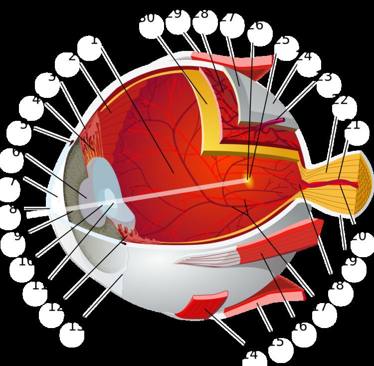
The vitreous body is the clear gel that fills the space between the lens and the retina of the eyeball of humans and other vertebrates. It is often referred to as the vitreous humour or simply "the vitreous".
Contents
Structure
The vitreous humour is a transparent, colorless, gelatinous mass that fills the space in the eye between the lens and the retina. It is present at birth. Produced by cells in the non-pigmented portion of the ciliary body, the vitreous humour is derived from embryonic mesenchyme cells, which degenerate after birth.
The vitreous humour is in contact with the retina and helps to keep it in place by pressing it against the choroid. It does not adhere to the retina, except at the optic nerve disc and the ora serrata (where the retina ends anteriorly), at the Wieger-band, the dorsal side of the lens. It is not connected at the macula, the area of the retina which provides finer detail and central vision.
Clinical
Unlike the fluid in the frontal parts of the eye (aqueous humour) which is continuously replenished, the gel in the vitreous chamber is stagnant. Therefore, if blood, cells or other byproducts of inflammation get into the vitreous, they will remain there unless removed surgically. These are known as floaters. If the vitreous pulls away from the retina, it is known as a vitreous detachment. As the human body ages, the vitreous often liquefies and may collapse. This is more likely to occur, and occurs much earlier, in eyes that are nearsighted (myopia). It can also occur after injuries to the eye or inflammation in the eye (uveitis).
The collagen fibres of the vitreous are held apart by electrical charges. With aging, these charges tend to reduce, and the fibres may clump together. Similarly, the gel may liquefy, a condition known as synaeresis, allowing cells and other organic clusters to float freely within the vitreous humour. These allow floaters which are perceived in the visual field as spots or fibrous strands. Floaters are generally harmless, but the sudden onset of recurring floaters may signify a posterior vitreous detachment (PVD) or other diseases of the eye.
The metabolic exchange and equilibration between systemic circulation and vitreous humour is so slow that vitreous humour is sometimes the fluid of choice for postmortem analysis of glucose levels or substances which would be more rapidly diffused, degraded, excreted or metabolized from the general circulation.
Forensic
After death, the vitreous resists putrifaction longer than other body fluids. The vitreous potassium concentration rises so predictably within the hours, days and weeks after death, that vitreous potassium levels are frequently used to estimate the time-of-death (Post-mortem interval) of a corpse.
Biochemical
Its composition is similar to that of the cornea, but the vitreous contains very few cells. It is composed mostly of phagocytes, which remove unwanted cellular debris in the visual field, and hyalocytes, which turn over the hyaluronan.
The vitreous humour contains no blood vessels, and 98–99% of its volume is water (as opposed to only 75% in the cornea). In addition to water, the vitreous consists of salts, sugars, vitrosin (a type of collagen), a network of collagen type II fibrils with glycosaminoglycan, hyaluronan, opticin, and a wide array of proteins. Despite having little solid matter, the fluid is substantial enough to fill the eye and give it its spherical shape. The lens, on the other hand, is tightly packed with cells. The vitreous humour has a viscosity two to four times that of water, giving it a gelatinous consistency. It has a refractive index of 1.336.
Anatomical
The vitreous has many anatomical landmarks, including the hyaloid membrane, Berger's space, space of Erggelet, Wieger's ligament, Cloquet's canal and the space of Martegiani.
Surface features:
Internal structures of the vitreous
Named tracts
Aging