
Entrez 3480 | Ensembl ENSG00000140443 | |
 | ||
Aliases IGF1R, CD221, IGFIR, IGFR, JTK13, insulin like growth factor 1 receptor, Insulin-like growth factor 1,IGF-1R External IDs OMIM: 147370 MGI: 96433 HomoloGene: 30997 GeneCards: IGF1R | ||
The insulin-like growth factor 1 (IGF-1) receptor is a protein found on the surface of human cells. It is a transmembrane receptor that is activated by a hormone called insulin-like growth factor 1 (IGF-1) and by a related hormone called IGF-2. It belongs to the large class of tyrosine kinase receptors. This receptor mediates the effects of IGF-1, which is a polypeptide protein hormone similar in molecular structure to insulin. IGF-1 plays an important role in growth and continues to have anabolic effects in adults - meaning that it can induce hypertrophy of skeletal muscle and other target tissues. Mice lacking the IGF-1 receptor die late in development, and show a dramatic reduction in body mass, testifying to the strong growth-promoting effect of this receptor. Mice carrying only one functional copy of IGF-1R are normal, but exhibit a ~15% decrease in body mass.
Contents
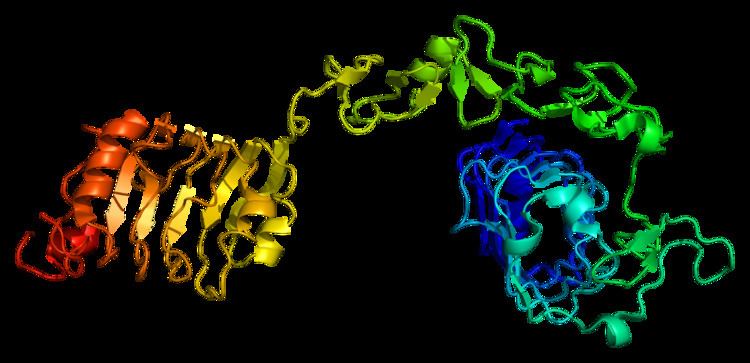
Structure
Two alpha subunits and two beta subunits make up the IGF-1 receptor. Both the α and β subunits are synthesized from a single mRNA precursor. The precursor is then glycosylated, proteolytically cleaved, and crosslinked by cysteine bonds to form a functional transmembrane αβ chain. The α chains are located extracellularly, while the β subunit spans the membrane and is responsible for intracellular signal transduction upon ligand stimulation. The mature IGF-1R has a molecular weight of approximately 320 kDa.citation? The receptor is a member of a family which consists of the insulin receptor and the IGF-2R (and their respective ligands IGF-1 and IGF-2), along with several IGF-binding proteins.
IGF-1R and the insulin receptor both have a binding site for ATP, which is used to provide the phosphates for autophosphorylation (see below). There is a 60% homology between IGF-1R and the insulin receptor.
In response to ligand binding, the α chains induce the tyrosine autophosphorylation of the β chains. This event triggers a cascade of intracellular signaling that, while cell type-specific, often promotes cell survival and cell proliferation. The structures of the autophosphorylation complexes of tyrosine residues 1165 and 1166 have been identified within crystals of the IGF1R kinase domain.
Family members
Tyrosine kinase receptors, including the IGF-1 receptor, mediate their activity by causing the addition of a phosphate groups to particular tyrosines on certain proteins within a cell. This addition of phosphate induces what are called "cell signaling" cascades - and the usual result of activation of the IGF-1 receptor is survival and proliferation in mitosis-competent cells, and growth (hypertrophy) in tissues such as skeletal muscle and cardiac muscle.
During embryonic development, the IGF-1R pathway is involved with the developing limb buds.
The IGFR signalling pathway is of critical importance during normal development of mammary gland tissue during pregnancy and lactation. During pregnancy, there is intense proliferation of epithelial cells which form the duct and gland tissue. Following weaning, the cells undergo apoptosis and all the tissue is destroyed. Several growth factors and hormones are involved in this overall process, and IGF-1R is believed to have roles in the differentiation of the cells and a key role in inhibiting apoptosis until weaning is complete.
Role in cancer
The IGF-1R is implicated in several cancers, including breast, prostate, and lung cancers. In some instances its anti-apoptotic properties allow cancerous cells to resist the cytotoxic properties of chemotherapeutic drugs or radiotherapy. In breast cancer, where EGFR inhibitors such as erlotinib are being used to inhibit the EGFR signaling pathway, IGF-1R confers resistance by forming one half of a heterodimer (see the description of EGFR signal transduction in the erlotinib page), allowing EGFR signaling to resume in the presence of a suitable inhibitor. This process is referred to as crosstalk between EGFR and IGF-1R. It is further implicated in breast cancer by increasing the metastatic potential of the original tumour by conferring the ability to promote vascularisation.
Increased levels of the IGF-IR are expressed in the majority of primary and metastatic prostate cancer patient tumors. Evidence suggests that IGF-IR signaling is required for survival and growth when prostate cancer cells progress to androgen independence. In addition, when immortalized prostate cancer cells mimicking advanced disease are treated with the IGF-1R ligand, IGF-1, the cells become more motile. Members of the IGF receptor family and their ligands also seem to be involved in the carcinogenesis of mammary tumors of dogs. IGF1R is amplified in several cancer types based on analysis of TCGA data, and gene amplification could be one mechanism for overexpression of IGF1R in cancer.
Role in insulin signaling
IGF-1 binds to at least two cell surface receptors: the IGF1 Receptor (IGFR), and the insulin receptor. The IGF-1 receptor seems to be the "physiologic" receptor - it binds IGF-1 at significantly higher affinity than it binds insulin. Like the insulin receptor, the IGF-1 receptor is a receptor tyrosine kinase - meaning it signals by causing the addition of a phosphate molecule on particular tyrosines. IGF-1 activates the insulin receptor at approximately 0.1x the potency of insulin. Part of this signaling may be via IGF1R/insulin receptor heterodimers (the reason for the confusion is that binding studies show that IGF1 binds the insulin receptor 100-fold less well than insulin, yet that does not correlate with the actual potency of IGF1 in vivo at inducing phosphorylation of the insulin receptor, and hypoglycemia).
Effects of aging
Studies in female mice have shown that both supraoptic nucleus (SON) and paraventricular nucleus (PVN) lose approximately one-third of IGF-1R immunoreactive cells with normal aging. Also, old calorically restricted (CR) mice lost higher numbers of IGF-1R non-immunoreactive cells while maintaining similar counts of IGF-1R immunoreactive cells in comparison to old-Al mice. Consequently, old-CR mice show a higher percentage of IGF-1R immunoreactive cells, reflecting increased hypothalamic sensitivity to IGF-1 in comparison to normally aging mice.
Role in craniosynostosis
Mutations in IGF1R have been associated with craniosynostosis.
Gene inactivation/deletion
Deletion of the IGF-1 receptor gene in mice results in lethality during early embryonic development, and for this reason, IGF-1 insensitivity, unlike the case of growth hormone (GH) insensitivity (Laron syndrome), is not observed in the human population.
Inhibitors
Due to the similarity of the structures of IGF-1R and the insulin receptor (IR), especially in the regions of the ATP binding site and tyrosine kinase regions, synthesising selective inhibitors of IGF-1R is difficult. Prominent in current research are three main classes of inhibitor:
- Tyrphostins such as AG538 and AG1024. These are in early pre-clinical testing. They are not thought to be ATP-competitive, although they are when used in EGFR as described in QSAR studies. These show some selectivity towards IGF-1R over IR.
- Pyrrolo(2,3-d)-pyrimidine derivatives such as NVP-AEW541, invented by Novartis, which show far greater (100 fold) selectivity towards IGF-1R over IR.
- Monoclonal antibodies are probably the most specific and promising therapeutic compounds. Those currently undergoing trials include figitumumab.
Interactions
Insulin-like growth factor 1 receptor has been shown to interact with:
Regulation
There is evidence to suggest that IGF1R is negatively regulated by the microRNA miR-7.