
Specialty ophthalmology ICD-9-CM 364.41 MedlinePlus 001021 | ICD-10 H21.0 DiseasesDB 31299 eMedicine oph/765 | |
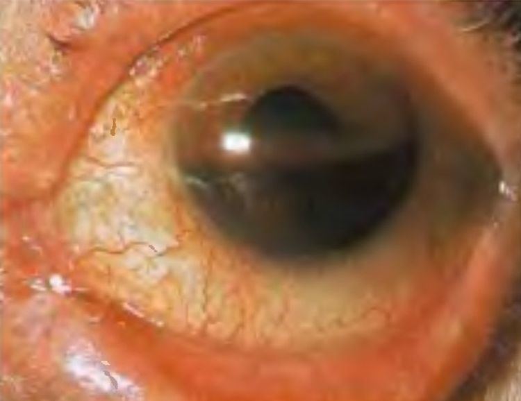 | ||
Hyphema (or hyphaema, see spelling differences) is blood in the front (anterior) chamber of the eye. It may appear as a reddish tinge, or it may appear as a small pool of blood at the bottom of the iris or in the cornea.
Contents
Epidemiology
As of 2012, the rate of hyphemas in the United States are about 20 cases per 100,000 people annually.
Causes
Hyphemas are frequently caused by injury, and may partially or completely block vision. The most common causes of hyphema are intraocular surgery, blunt trauma, and lacerating trauma. Hyphemas may also occur spontaneously, without any inciting trauma. Spontaneous hyphemas are usually caused by the abnormal growth of blood vessels (neovascularization), tumors of the eye (retinoblastoma or iris melanoma), uveitis, or vascular anomalies (juvenile xanthogranuloma). Additional causes of spontaneous hyphema include: rubeosis iridis, myotonic dystrophy, leukemia, hemophilia, and von Willebrand disease. Conditions or medications that cause thinning of the blood, such as aspirin, warfarin, or drinking alcohol may also cause hyphema.
Prognosis
Hyphemas require urgent assessment by an optometrist or ophthalmologist as they may result in permanent visual impairment.
A long-standing hyphema may result in hemosiderosis and heterochromia. Blood accumulation may also cause an elevation of the intraocular pressure. On average, the increased pressure in the eye remains for six days before dropping. Most uncomplicated hyphemas resolve within 5–6 days.
Treatment
The main goals of treatment are to decrease the risk of rebleeding within the eye, corneal blood staining, and atrophy of the optic nerve. Small hyphemas can usually be treated on an outpatient basis. Most treatment plans consist of elevating the head at night, wearing a patch and shield, and controlling any increase in intraocular pressure. Surgery may be necessary for non-resolving hyphemas, or hyphaemas that are associated with high pressure that does not respond to medication. Surgery can be effective for cleaning out the anterior chamber and preventing corneal blood staining.
Elevation of the head of the bed by approximately 45 degrees (so that the hyphema can settle out inferiorly and avoid obstruction of vision, as well as to facilitate resolution). Bed rest may be considered, although evidence suggests that it does not improve outcomes. Wearing of an eye shield at night time (to prevent accidental rubbing of the eyes during sleep, which can precipitate a rebleed). An eye patch should be worn throughout the day to protect the injured eye.
If pain management is necessary, acetaminophen can be used. Aspirin and ibuprofen should be avoided, because they thin the blood and increase the risk of a rebleed. Sedation is not usually necessary for patients with hyphema. It is controversial amongst eye practitioners whether a steroid medication or a dilating drop (mydriatic) should be used in treatment of hyphema. Steroids aim to reduce the amount of inflammation, but also cause side effects. Dilating drops aim to increase comfort from the traumatized iris as well as reduce bleeding, but can also cause the pupil to be fixed in a dilated state via posterior synechiae (adhesions).
Aminocaproic or tranexamic acids are often prescribed for hyphema. Although these medications actually cause hyphemas to take longer to clear, they reduce the risk of rebleeding and its associated complications. Tranexamic and aminocaproic acids inhibit the conversion of plasminogen to plasmin, plasmin being the agent of fibrin breakdown in blood clots. Keeping the clots intact allows time for the vessels to heal properly and avert a secondary bleed.
Complications
While the vast majority of hyphemas resolve on their own without issue, sometimes complications occur. Traumatic hyphema may lead to increased intraocular pressure, peripheral anterior synechiae, atrophy of the optic nerve, staining of the cornea with blood, re-bleeding, and impaired accommodation.
Secondary hemorrhage, or rebleeding of the hyphema, is thought to worsen outcomes in terms of visual function. Rebleeding occurs in 4-35% of hyphema cases and is a risk factor for glaucoma.