
Entrez 6517 | Ensembl ENSG00000181856 | |
 | ||
Aliases SLC2A4, GLUT4, solute carrier family 2 member 4 External IDs OMIM: 138190 MGI: 95758 HomoloGene: 74381 GeneCards: SLC2A4 | ||
Glucose transporter type 4, also known as GLUT4, is a protein encoded, in humans, by the GLUT4 gene. GLUT4 is the insulin-regulated glucose transporter found primarily in adipose tissues and striated muscle (skeletal and cardiac). The first evidence for this distinct glucose transport protein was provided by David James in 1988. The gene that encodes GLUT4 was cloned and mapped in 1989.
Contents
- Tissue distribution
- Insulin
- Muscle contraction
- Muscle stretching
- Interactions
- Interactive pathway map
- References
Recent reports demonstrated the presence of GLUT4 gene in central nervous system such as the hippocampus. Moreover, impairment in insulin-stimulated trafficking of GLUT4 in the hippocampus result in decreased metabolic activities and plasticity of hippocampal neurons, which leads to depressive like behaviour and cognitive dysfunction.
Tissue distribution
GLUT4 is primarily found in:
Insulin
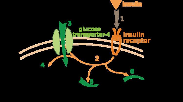
Under conditions of low insulin, most GLUT4 is sequestered in intracellular vesicles in muscle and fat cells. Insulin induces a rapid increase in the uptake of glucose by inducing the translocation of GLUT4 from these vesicles to the plasma membrane. As the vesicles fuse with the plasma membrane, GLUT4 transporters are inserted and become available for transporting glucose, and glucose absorption increases. Insulin’s actions are effectively abolished in the genetically engineered muscle insulin receptor knock‐out (MIRKO) mouse because they have a complete lack of the insulin‐sensitive glucose transport protein, GLUT4, in muscle, so insulin has no effect on glucose uptake. Fascinatingly, this is of little or no consequence to the animal that does not have fasting hyperglycaemia or diabetes.
Insulin binds to the insulin receptor in its dimeric form and activates the receptor's tyrosine-kinase domain. The receptor then phosphorylates and subsequently recruits Insulin Receptor Substrate or IRS-1, which in turn binds the enzyme PI-3 kinase through the binding of the enzyme's SH2 domain to the pTyr of IRS. PI-3 kinase converts the membrane lipid PIP2 to PIP3. PIP3 is specifically recognized by the PH domains of PKB (protein kinase B) or AKT, and also for PDK1 which, being localized together with PKB, can phosphorylate and activate PKB. Once phosphorylated, PKB is in its active form and phosphorylates TBC1D4, which inhibits the GAP domain or the GTPase-activating domain associated with TBC1D4, allowing for Rab protein to change from its GDP to GTP bound state. Inhibition of the GTPase-activating domain leaves proteins next in the cascade in their active form and stimulates GLUT4 to be expressed on the plasma membrane. RAC1 is a GTPase which is also activated by insulin. Rac1 stimulates reorganization of the cortical Actin cytoskeleton which allows for the GLUT4 vesicles to be inserted into the plasma membrane. RAC1 Knockout mouse have reduced glucose uptake in muscle.
At the cell surface, GLUT4 permits the facilitated diffusion of circulating glucose down its concentration gradient into muscle and fat cells. Once within cells, glucose is rapidly phosphorylated by glucokinase in the liver and hexokinase in other tissues to form glucose-6-phosphate, which then enters glycolysis or is polymerized into glycogen. Glucose-6-phosphate cannot diffuse back out of cells, which also serves to maintain the concentration gradient for glucose to passively enter cells.
Knockout mice that are heterozygous for GLUT4 develop insulin resistance in their muscles as well as diabetes.
Muscle contraction
Muscle contraction stimulates muscle cells to translocate GLUT4 receptors to their surfaces. This is especially true in cardiac muscle, where continuous contraction can be relied upon; but is observed to a lesser extent in skeletal muscle. In skeletal muscle, muscle contraction increase GLUT4 translocation several fold and this is likely regulated by RAC1 and AMP-activated protein kinase.
Muscle stretching
Muscle stretching also stimulate GLUT4 translocation and glucose uptake in rodent muscle via RAC1.
Interactions
GLUT4 has been shown to interact with death-associated protein 6.
Interactive pathway map
Click on genes, proteins and metabolites below to link to respective articles.