
Dorlands/Elsevier l_09/12493077 FMA 57077 | TA A03.1.07.007 | |
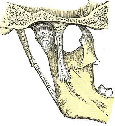 | ||
Latin ligamentum sphenomandibulare | ||
The sphenomandibular ligament (internal lateral ligament) is a flat, thin band which is attached superiorly to the spina angularis (spine) of the sphenoid bone, and, becoming broader as it descends, is fixed to the lingula of the mandibular foramen. The function of the sphenomandibular ligament is to limit distension of the mandible in an inferior direction. It is slack when the temporomandibular joint (TMJ) is in closed position. It is taut as the condyle of the mandible is in front of the temporomandibular ligament.
Lateral pterygoid and the auriculotemporal nerve are lateral relations, the chorda tympani nerve lies medial near its upper end and medial pterygoid is an inferomedial relation. The sphenomandibular ligament is separated from the neck of the mandible below lateral pterygoid by the maxillary artery and from the ramus of the mandible by the inferior alveolar vessels and nerve and a parotid lobule.
The ligament is derived from Meckel's cartilage.