
TA A02.5.11.001 | FMA 24496 | |
 | ||
Latin Calcaneus, Calcaneum, Os calcis MeSH A02.835.232.043.300.710.300 | ||
In humans, the calcaneus (/kælˈkeɪniːəs/; from the Latin calcaneus or calcaneum, meaning heel) or heel bone is a bone of the tarsus of the foot which constitutes the heel. In some other animals, it is the point of the hock.
Contents
Structure
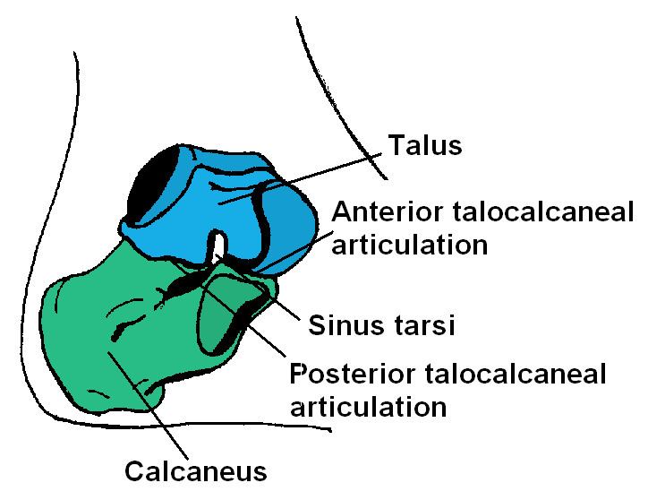
In humans, the calcaneus is the largest of the tarsal bones and the largest bone of the foot. The talus bone, calcaneus, and navicular bone are considered the proximal row of tarsal bones. In the calcaneus, several important structures can be distinguished:
The half of the bone closest to the heel is the calcaneal tubercle. On its lower edge on either side are its lateral and medial processes (serving as the origins of the abductor hallucis and abductor digiti minimi). The Achilles tendon is inserted into a roughened area on its superior side, the cuboid bone articulates with its anterior side, and on its superior side are three articular surfaces for the articulation with the talus bone. Between these superior articulations and the equivalents on the talus is the tarsal sinus (a canal occupied by the interosseous talocalcaneal ligament). At the upper and forepart of the medial surface of the calcaneus, below the middle talar facet, there is a horizontal eminence, the talar shelf (also sustentaculum tali), which gives attachment to the plantar calcaneonavicular (spring) ligament, tibiocalcaneal ligament, and medial talocalcaneal ligament. This eminence is concave above, and articulates with the middle calcaneal articular surface of the talus; below, it is grooved for the tendon of the flexor hallucis longus; its anterior margin gives attachment to the plantar calcaneonavicular ligament, and its medial margin to a part of the deltoid ligament of the ankle-joint.
On the lateral side is commonly a tubercle called the calcaneal tubercle (or trochlear process). This is a raised projection located between the tendons of the peroneus longus and brevis. It separates the two oblique grooves of the lateral surface of the calcaneus (for the tendons of the peroneal muscles).
Its chief anatomical significance is as a point of divergence of the previously common pathway shared by the distal tendons of peroneus longus and peroneus brevis en route to their distinct respective attachment sites.
The calcaneus is part of two joints: the proximal intertarsal joint and the talocalcaneal joint. The point of the calcaneus is covered by the calcanean bursa.
Development
In the calcaneus, an ossification center, is developed during the 4th–7th week of fetal development.
Function
Three muscles attach to the calcaneus: the gastrocnemius, soleus, and plantaris. These muscles are part of the posterior compartment of the leg and aid in walking, running and jumping. Their specific functions include plantarflexion of the foot, flexion of the knee, and steadying the leg on the ankle during standing.
Clinical significance
Normally the tibia sits vertically above the calcaneus (pes rectus). If the calcaneal axis between these two bones is turned medially the foot is in an everted position (pes valgus), and if it is turned laterally the foot is in an inverted position (pes varus).
Disease
The talar shelf is typically involved in subtalar or talocalcaneal tarsal coalition.